Download
1 / 17
250 likes | 950 Views
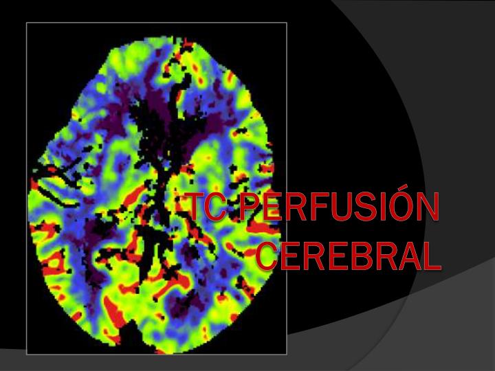
TC PERFUSIÓN CEREBRAL. TC PERFUSIÓN CEREBRAL. PRINCIPIOS BÁSICOS. La perfusión del tejido cerebral normal se mantiene dentro de un rango muy estrecho por la autorregulación de la vascularización cerebral Flujo sanguíneo normal en Sustancia Gris de 50-60 mL /100g/min

E N D
TC PERFUSIÓN CEREBRAL PRINCIPIOS BÁSICOS • La perfusión del tejido cerebral normal se mantiene dentro de un rango muy estrecho por la autorregulación de la vascularización cerebral • Flujo sanguíneo normal en Sustancia Gris de 50-60 mL/100g/min • La supervivencia del tejido isquémico tras la oclusión arterial depende de la existencia de un aporte colateral mínimo a través de anastomosis leptomeníngeas.
FLUJO SANGUÍNEO CEREBRAL (CBF) TC PERFUSIÓN CEREBRAL CAMBIOS EN FUNCIÓN Y SUPERVIVENCIA DEL TEJIDO ISQUÉMICO: MODELO TRICOMPARTIMENTAL
TC PERFUSIÓN CEREBRAL PRINCIPIOS BÁSICOS
TC PERFUSIÓN CEREBRAL PRINCIPIOS BÁSICOS CURVAS DE ATENUACIÓN / TIEMPO/ UNIDAD VOXEL UH Pendiente máxima (CBF) Área bajo la curva (CBV) TTP Medidas de Captación Captación Máxima Tiempo a inicio Tiempo (s) 0 10 20 30
TC perfusión: Técnica TC cerebral en vacío Se seleccionan dos secciones localizadas Sobre núcleos básales En centro semioval Se administra 50 ml contraste (4-5 ml/s.) Se obtienen 20 cortes axiales consecutivos de 12 mm de grosor, a 1 corte/seg. Se determinan los cambios en la atenuación cerebral
CURVAS DE CAPTACIÓN TC PERFUSIÓN TER.TEMPOROPARIETAL ACM 4 5 2 3 SENO SAGITAL SUPERIOR TERRITORIO ANTERIOR ACM 1 PARÉNQUIMA CEREBRAL NORMAL ÁREA NO PERFUNDIDA
TC perfusión En el ictus el tejido en penumbra Aumenta el TTM Disminuye el CBF (>60%) Aumenta el CBV (>80-100%) El tejido necrótico Aumenta el TTM Disminuye el CBF (<30%) Disminuye el CBV (< 40%)
TC perfusión Los mapas de TTM son los mas sensiblespara diferenciar ictus de penumbra. Los mapas de CBF y CBV son mas específicos. El mismatch CBF-CBV indica el área en penumbra Eastwood et al, Radiology 2002, 222:227-36
CONCLUSIONES TC PERFUSIÓN CEREBRAL • Amplia disponibilidad y rápida realización. • TC helicoidal estándar. • Mapas de perfusión en corto periodo de tiempo en estación de trabajo apropiada. • Evaluación cuantitativa y cualitativa: • CBV (Volumen Sanguíneo Cerebral) • CBF (Flujo Sanguíneo Cerebral) • TTP (Tiempo de Pico) • MTT (Tiempo Medio de Tránsito)