Download
1 / 69
880 likes | 1.58k Views
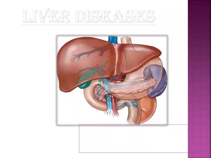
LIVER DISEASES. Physiology of liver. Liver is the largest organ of the body. Sets in the right side of the belly. Weighs between 1.0-2.5 kg. Heavier in males than females. It is reddish in color & feel rubbery to touch. Have two sections-right & left lobe.

E N D
Physiology of liver • Liver is the largest organ of the body. • Sets in the right side of the belly. • Weighs between 1.0-2.5 kg. • Heavier in males than females. • It is reddish in color & feel rubbery to touch. • Have two sections-right & left lobe. • The right lobe is further divided into the interior and posterior segments
The externally visible falciform ligament divides the left lobe into the medial and lateral segments. • Liver is supplied with blood from two sources –the hepatic artery & portal vein. • Responsible for 25 %of the basal metabolism & intimately concerned with the metabolism of fats,protiens & CHO.
structure of liver • Composed of 50,000-100,000 lobules. • Each lobule consist of a central vein surrounded by tiny liver cells grouped in the sheet or bundle. • Small branch of hepatic vein extends through their centre. . • Space b/w these cells contain sinusoids (blood vessels that are irregular in shape, incomplete in size &wider than blood capillaries .)
Blood supply • the liver receives blood containing oxygen from the heart. This blood enters the liver through the hepatic artery. • The liver also receives blood filled with nutrients, or digested food particles, from the small intestine. This blood enters the liver through the portal vein. • In the liver, the hepatic artery and the portal vein branch into a network of tiny blood vessels that empty into the sinusoids. • The liver cells absorb nutrients and oxygen from the blood as it flows through the sinusoids. At the same time, they secrete sugar, vitamins, minerals, and other substances into the blood. • The sinusoids drain into the central veins, which join to form the hepatic vein. Blood leaves the liver through the hepatic vein. • Each lobule also contains bile capillaries, tiny tubes that carry the bile secreted by the liver cells. The bile capillaries join to form bile ducts, which carry bile out of the liver. Soon after leaving the liver, the bile ducts join together, forming the hepatic duct. • Excess bile flows into the gall bladder, where it is stored for later use.
Functions of liver • Secretion of bile • Storage of glycogen • Metabolism of fats • Deamination of amino acids • Production of the plasma protiens. • Storage & transport of vitamins & minerals. • Storage of iron . • Production of clotting factors . • Production of heat . • Detoxification. • Acts as filter
Role of branched chain amino acids & aromatic amino acids in liver disease • major hypothesis for the pathogenesis of portal systemic encephalopathy has been termed the altered neurotransmitter Theory. • A plasma amino acid imbalance existsin ESLD in which the branched-chain amino acids( BGAAs) valine, leucine, and isoleucine are decreased, and aromatic amino acids (AAAs) tryptophan, phenylalanine, and tryrosine,plus methionine, glutamine, asparagine, and histidine are increased. • The BCAAs furnish as much as 30% of energy requirements for skeletal muscle, heart, and brain when gluconeogenesis and ketogenesis are depressed . • This causes serum BCAA levels to fall. At the same time, plasma AAA’S and methionine are released into circulation by muscle proteolysis, but the synthesis into protein and liver clearance of AAAs is depressed . • This changes the plasma molar ratio of BCAAs to AAA’S and may contribute to the development of hepatic encephalopathy. • AAAs may limit the cerebral uptake of BCAA because they compete for carrier-mediated transport at the blood-brain barrier. ..
1.Aspartate aminotransferase (AST or SGOT) • Located in cytosol and mitochondria of hepatocyte also in cardiac and skeletal muscle, brain, pancreas, kidney and leukocytes. • An enzyme similar to ALT but less specific for liver disease. • Levels of AST are increased with liver damage or death of hepatocytes. • In some cases viz alcoholic hepatitis or shock liv the elevation in serum AST level may be, higher than the elevation in serum ALT level. • AST Normal range – 2-40 U/L.
2.Alanine aminotransferase (ALT or SGPT) • It is located in cytosol of hepatocytes found in several other body tissues. • It is highest in liver. • ALT level is increased in case of liver cell death, caused due to, Hepatitis, any shock, injury or excess alcohol or drug toxicity. • Normal range – 2-40 U/L
3.Serum alkaline phosphatase • An enzyme that is widely distributed in liver, bone, placenta, intestine, kidney, leukocytes. • It is mainly bound to canalicular membranes of liver produced in the bile ducts. • This enzyme activity can be increased with bone disorders, pregnancy , normal growth & some malignancies. • 40 - 129 U/L
4.bilirubin • Bilirubin is a yellow breakdown product of the normal heme catabolism . • It is excreted in bile & urine and elevated levels may indicate certain diseases. • It is responsible for the yellow color of the bruises ,urine & brown color of feces & yellow discoloration of skin.
functions • Bilirubin is created by the activity of biliverdin reductase on biliverdin, a green tetrapyrrolic bile pigment that is also a product of heme catabolism. Bilirubin, when oxidized, reverts to become biliverdin once again. This cycle, in addition to the demonstration of the potent antioxidant activity of bilirubin, has led to the hypothesis that bilirubin's main physiologic role is as a cellular antioxidant.
Types of bilirubin • Unconjugated bilirubin (indirect) • Erythrocytes (red blood cells) generated in the bone marrow are disposed of in the spleen when they get old or damaged. This releases hemoglobin, which is broken down to heme as the globin parts are turned into amino acids. The heme is then turned into unconjugated bilirubin in the reticuloendothelial cells of the spleen. This unconjugated bilirubin is not soluble in water, due to intramolecular hydrogen bonding. It is then bound to albumin and sent to the liver • Conjugated bilirubin(direct) • In the liver it is conjugated with glucuronic acid by the enzyme glucuronyltransferase, making it soluble in water. Much of it goes into the bile and thus out into the small intestine. However 95% of the secreted bile is reabsorbed by the small intestine. This bile is then resecreted by the liver into the small intestine. This process is known as enterohepatic circulation. About half of the conjugated bilirubin remaining in the large intestine(about 5% of what was originally secreted) is metabolized by colonic bacteria to urobilinogen, which can be further metabolized to stercobilinogen, and finally oxidized to stercobilin. Stercobilin gives feces its brown color
Blood tests • Bilirubin is broken down by light. Tubes containing blood or (especially) serum to be used in bilirubin assays should be protected from illumination. • Total bilirubin ("TBIL") measures both BU and BC. Total and direct bilirubin levels can be measured from the blood, but indirect bilirubin is calculated from the total and direct bilirubin. • Indirect bilirubin is fat-soluble and direct bilirubin is water-soluble. • Bilirubin (in blood) is in two forms.
Hyperbilirubinemia • Hyperbilirubinemia results from a higher than normal level of bilirubin in the blood. • Mild rises in bilirubin may be caused by: • Hemolysis or increased breakdown of red blood cells • Gilbert's syndrome - a genetic disorder of bilirubin metabolism that can result in mild jaundice, found in about 5% of the population • Rotor syndrome: non-itching jaundice, with rise of bilirubin in the patient's serum, mainly of the conjugated type. • Moderate rise in bilirubin may be caused by: • Pharmaceutical drugs (especially antipsychotic, some sex hormones, and a wide range of other drugs) • Sulfonamides are contraindicated in infants less than 2 months old as they increase unconjugated bilirubin leading to kernicterus. • Hepatitis (levels may be moderate or high) • Chemotherapy • Biliary stricture (benign or malignant) • Very levels of bilirubin may be caused by: • Neonatal hyperbilirubinaemia, where the newborn's liver is not able to properly process the bilirubin causing jaundice • Unusually large bile duct obstruction, e.g. stone in common bile duct, tumour obstructing common bile duct etc. • Severe liver failure with cirrhosis (e.g. primary biliary cirrhosis • Choledocholithiasis (chronic or acute)presence of gallstone in the common bile duct
5.Gamma-glutamyl transpeptidase (GGT) • An enzyme associated with microsomes and plasma membranes in hepatocytes also present in kidney, pancreas, heart , brain. • An enzyme produced in the bile ducts that like alkaline phosphatase, may be elevated in the serum of patients with bile duct disease. • It is increased with liver disease, pulmonary disease, diabetes mellitus and during alcohol ingestion by many drugs.
Serum albumin • Main export protein synthesized in the liver and most imp. factor in plasma osmotic pressure maintenance, secreted in the blood. • Major protein circulating in blood stream. • Low serum albumin levels indicate poor liver function. • Serum albumin concentration is usually normal in chronic liver diseases until cirrhosis and liver damage is present. • Albumin – 3.4 - 4.8 g%. • Albumin levels can be low in conditions other than, liver disease like malnutrition. • Albumin is a better index of chronic liver disease. • Increased losses occur with protein losing enteropathy, nephrotic syndrome burns, GIT bleeding, exfoliative dermatitis.
Prothrombin time • Most blood coagulation factors are synthesized in the liver. • When liver function is severely abnormal, their synthesis and secretion into blood is decreased. • PT is a type of blood clotting test and is prolonged when blood concentration of some clotting factors made by liver are low. • In chronic liver disease, PT is not usually elevated until cirrhosis is present and liver damage is significant. • In acute liver disease, PT can be prolonged with severe liver damage. • Decreased synthesis of clotting factors increase prothrombin time and risk of bleeding.
Serum protein electrophoresis • Major protein in the serum are separated in an electric field and their concentrations are determined. • Four major types of serum proteins measured are albumin, alpha- globulins, beta- globulins and gamma- globulins. • Alpha and gamma globulins are synthesized in liver. Levels increase with chronic liver disease. • In cirrhosis, the albumin may be decreased and the gamma - globulins elevated. • Gamma- globulins can be significantly elevated in some types of autoimmune hepatitis also.
Platelet count • Platelets are the smallest of the blood cells, involved in blood clotting. • With liver disease the spleen becomes enlarged as blood flow through the liver is impeded. • This can lead to platelets being sequestered in the enlarged spleen. • The platelet count can be abnormal in many conditions other than liver disease.
gallbladder • It is a thin walled reservoir situated on the under surface of liver. • It can store about 40-50ml of bile. • It concentrates bile formed in the liver. • Stores it , until needed for digestion. • Interference with the flow of bile in gallbladder diseases may cause impaired fat digestion.
Functions of gall bladder • Acts as a reservoir for the storage of bile . • Mucosal lining of gall bladder reabsorbs the fluid & electrolytes ,thus concentrating the bile constituents. • Hormone cholecystokinin secreted by the duodenal walls, cause muscular walls of the gall bladder to contract and expel the bile. • Duodenal peristalsis inhibits the sphincter of oddi & cause it to relax & allow the bile to enter the duodenum.
bile • Bile is the external secretion of the liver . • Produced in the diluted form . • Concentrated later by the gall bladder to a viscid greenish fluid . • It consists of- • Water • Bile salts(sodium glycocholate &sodium taurocholate) • Bile acids • Bile pigment( bilirubin). • Cholesterol • Mucus
Functions • Bile acts to some extent as a surfactant, helping to emulsify the fats in the food. • Since bile increases the absorption of fats, it is an important part of the absorption of the fat-soluble vitamins, such as the vitamins D, E, K and A. • Bile serves also as the route of excretion for bilirubin, a by-product of red blood cells, recycled by the liver. • Bile salts also act as bactericides, destroying many of the microbes that may be present in the food. • Bile salt anions have a hydrophilic side and a hydrophobic side, and therefore tend to aggregate around droplets of fat (triglycerides and phospholipids) to form micelles, with the hydrophobic sides towards the fat and hydrophilic towards the outside.
Bile salts • have important Function in assisting the digestive action of the pancreatic enzyme . • helps in aiding the absorption of fats & fat soluble vitamins form the small intestine. • These salts by lowering the surface tension cause fat droplets to break up or emulsify into small droplets allowing fat digesting enzyme to work more efficiently. • They convert them to fatty acids & glycerol ,the form in which they are absorbed. • Bile salts do not appear in faeces as they are reabsorbed from the small intestine & returned to the liver.
Bile acids • These include • 40% cholic acid . • 40% chemodeoxycholic acid. • 18% deooxycholic acid . • 2% lithocholic acid. • Total bile acid pool is 2.4 g but it is absorbed & recirculated 6-8 times a day. Thus providing over 15- 20 gm of bile for digestion.
Bile pigments • Are derived from the breakdown of the haemoglobin of worn out RBC’s & give the bile its characteristic color. • The bile pigments are converted in the bowel to urobilinogen by bacterial action. • Some urobilinogen is reabsorbed into the blood & is excreted by the kidneys into the urine. • Exposure of urine to the air causes urobilinogen to be oxidized to urobilin. • In the faeces , urobilinogen is altered 7 oxidized to form stercobilin which gives the faeces a dark brown colour. • If there is an obstruction to the excretion of bile , bile pigments accumulate in the blood giving the skin ,a yellow color (jaundice).
Bile Storage • The liver is constantly secreting bile, up to 1 liter in a 24 hour period, but most of it is stored in the gallbladder. • This hollow organ can only hold 30 to 60 ml of bile and is able to store the large quantities of bile from the liver by concentrating it. • The gallbladder is able to achieve this by reabsorption of water, sodium, chloride and other electrolytes through its lining. • The other constituents of bile, like the bile salts, cholesterol, lecithin and bilirubin, stays in the gallbladder.
Bile Production • The liver cells (hepatocytes) produce bile which collects and drains into the hepatic duct. • From here it can enter the small intestine to act on fats by traveling down the common bile duct, or it can enter the gallbladder through the cystic duct, where it is stored. • The liver manufactures between 600ml to 1 liter of bile in a day. As bile travels down the ducts, the lining of these passages, secrete water, sodium and bicarbonate ions into the bile, thereby diluting it. • These additional substances help to neutralize the stomach acid which enters the duodenum with partially digested food (chyme) from the stomach.
diseases • Cholelithiases
Cholelithiasis • The formation of gallstones (calculi) in the absence of infection of the gallbladder is called cholelithiasis. • all gallstones form within the gallbladder • Gallstones that pass from the gallbladder into the common bile duct may remain there indefinitely without causing symptoms, or they may pass into the duodenum with or without symptoms. • Choledocholithiasis develops when stones slip into the bile ducts, producing obstruction, pain, and cramps. • If passage of bile into the duodenum is interrupted, cholecystitis can develop. In the absence of bile in the intestine, lipid absorption is impaired, and without bile pigments, stools become light in color. • If uncorrected, bile backup can result in jaundice and liver damage (secondary biliary cirrhosis). • Obstruction of the distal common bile duct can lead to pancreatitis if the pancreatic duct is blocked.
Types of stones • Most gallstones in people are unpigmented cholesterol stones composed primarily of cholesterol ,bilirubin and calcium salts. • Risk factors for cholesterol stone formation include female gender, pregnancy, older age, family history obesity diabetes mellitus, inflammatory bowel disease and drugs (lipid-lowering medications, oral contraceptives and estrogens). • Rapid weight loss (as with jejunoileal and gastric bypass and fasting or severe calorie restriction) is associated with a high incidence of biliary sludge and gallstone formation . • Bacteria may also play a role in gallstone formation. Low-grade chronic infections produce changes in the gallbladder mucosa, which affect its absorptive capabilities. Excess water or bile acid may be absorbed as a result. Cholesterol may then precipitate out and cause gallstone formation .
High dietary fat intake over a prolonged period may predispose a person to gallstone formation because of the constant stimulus to produce more cholesterol for bile synthesis required in fat digestion. • Pigmented stones typically consist of bilirubin polymers or calcium salts. They are associated with chronic hemolysis. • Risk factors associated with these stones are age, sickle cell anemia and thalassemia, biliary tract infection, cirrhosis, alcoholism and long-term PN( parenteral nutrition).
Medical management • treatment of gallstone disease includes cholecystectomy, especially if the stones are numerous, large, or calcified. • cholecystectomy may be done as a traditional open laparotomy or as a less invasive laparoscopic procedure. • Chemical dissolution with the administration of bile salt with other acids are used but very less. • Patients with gallstones that have migrated into the bile ducts may be candidates for endoscopic retrograde cholangiopancreatography techniques
Nutrition therapy • No specific dietary treatment is available to prevent cholelithiasism in susceptible persons. • Nutrition-related factors include obesity ,severe fasting and these should be corrected when possible. • In cholecystitis, dietary treatment includes a low-fat diet to prevent gallbladder contractions. • After surgical removal of the gallbladder, oral feedings are usually resumed with the return of bowel movement. • after that the diet can be advanced to a regular diet as tolerated. • In the absence of the gallbladder, bile is secreted directly by the liver into the intestine.
Cholecystitis • Inflammation of the gallbladder is known as cholecystitis. • it may be chronic or acute. It is usually caused by gallstones obstructing the bile ducts , leading to the backup of bile. • Bilirubin, the main bile pigment, gives bile its greenish color. • When biliary tract obstruction prevents bile from reaching the intestine, it backs up and returns to the circulation.
Acute cholecystitis • Due to infection of gall bladder. • It occurs in association with obstruction to the cystic duct or neck of the gall bladder. • Gall stones are the cause of obstruction • The walls of the gallbladder become inflamed and distended and infection can occur. • During such episodes, the patient experiences upper quadrant abdominal pain accompanied by nausea, vomiting, and flatulence.
Nutritional therapy in acute cholecystitis • In an acute attack, oral feedings are discontinued. Parenteral nutrition may be indicated if the patient is malnourished. • When feedings are resumed, a low-fat diet is recommended to decrease gallbladder stimulation • Patient should be kept in bed & given analgesics. • Large quantities of fluids should be drunk. • For acute cases, an entirely fluid of at least 2 -3 litres daily given in small feeds at hourly or two hourly intervals is advisable for few days.
Chronic cholecystitis • Chronic cholecystitis is long-standing inflammation of the gallbladder. • It is caused by repeated mild attacks of acute cholecystitis. • This leads to thickening of the walls of the gallbladder. • The gallbladder begins to shrink and eventually loses the ability to perform its function: concentrating and storing bile. • Eating foods that are high in fat may aggravate the symptoms of cholecystitis because bile is needed to digest such foods. • Chronic cholecystitis occurs more often in women than in men and the incidence increases after the age of 40. • Risk factors include the presence of gallstones and a history of acute cholecystitis.