Download
1 / 2
20 likes | 144 Views
d. Methylation of Plk4 in Cirrhotic Adjacent Liver Tissue. -135 bp. -150 bp. -150 bp. -235 bp. b. c. Methylation of Plk1 and Plk4 in Human HCC Tumours. Controls. Cirrhotic adjacent. Controls. Methylation of Plks in Normal Human Liver Samples.

E N D
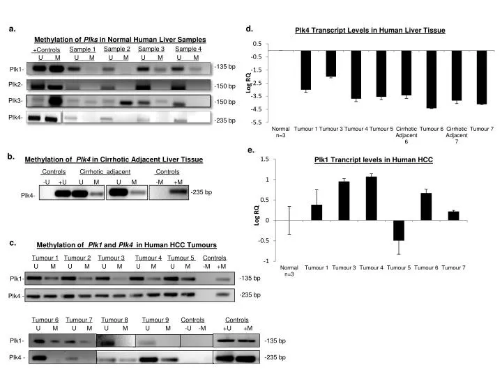
d. Methylation of Plk4 in Cirrhotic Adjacent Liver Tissue -135 bp -150 bp -150 bp -235 bp b. c. Methylation of Plk1 and Plk4 in Human HCC Tumours Controls Cirrhotic adjacent Controls Methylation of Plks in Normal Human Liver Samples -U +U U M U M -M +M a. Sample 2 Sample 3 Sample 4 Sample 1 +Controls -235 bp Plk4- U MU MU MU MU M Plk1- Plk2- Plk3- Plk4- Tumour 1Tumour 2Tumour 3Tumour 4Tumour 5 Controls U M U M U M U M U M -M +M Plk1- -135 bp Tumour 6Tumour 7Tumour 8Tumour 9ControlsControls Plk4 - U M U M U M U M -U -M +U +M -235 bp Plk1- -135 bp -235 bp Plk4 -
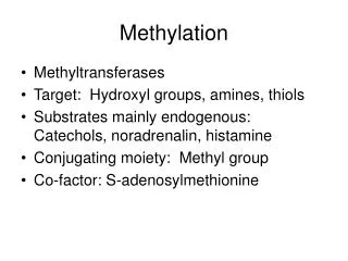
Additional file 2Plk4 CpG island methylation and expression levels in human liver samples. (a) Plk methylation status of genomic DNA extracted from normal human liver tissue was determined by MSP. Fully methylated human genomic DNA (NEB) was used as a positive control for methylation. The negative PCR control lacked DNA template. U=unmethylated, M=methylated (b) In contrast the analysis of cirrhotic adjacent tissue by MSP displayed increased levels of Plk4 methylation suggesting that methylation of Plk4 may be an early event in the transition of cirrhosis to HCC. (c) Shown is representative image of both the Plk4 and Plk1 methylation status in human HCC tumour samples as determined by MSP. Only two of the samples had hypomethylation within the Plk1 promoter region, suggesting that loss of Plk1 methylation may be a later event. U=unmethylated, M=methylated (d) Human HCC samples were analysed for Plk expression by qPCR. There was more than a 20 fold decrease in Plk4 transcript levels in tumours in comparison to normal liver tissue, while (e) Plk1 transcript levels were elevated in several of the tumour samples compared to the control. RQ values were normalized to the level of Plk4 transcripts in normal livers. Transcript levels for tumours 2, 8, and 9 were not assessed as they were archival specimens with poor quality RNA. Human GAPDH was used as an internal control.