Download

1 / 12
120 likes | 260 Views
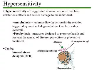
IgE mediated hypersensitivity. Allergens. Most allergic IgE responses occur on mucous membrane surfaces in response to allergens that enter the body by either inhalation or ingestion

E N D
Allergens • Most allergic IgE responses occur on mucous membrane surfaces in response to allergens that enter the body by either inhalation or ingestion • allergens are small proteins or protein-bound substances having a molecular weight between 15,000 and 40,000, • It appears that allergenicity is a consequence of a complex series of interactions involving not only the allergen but also the • dose, • the sensitizing route, sometimes • an adjuvant, and—most important • the genetic constitution of the recipient
Effector mechanisms in lgE-mediated allergic reactions • Mast-cell activation has different effects on different tissues • Mast cells line external mucosal surfaces and serve to alert the immune system to local infection • inflammatory reactions by secreting pharmacological mediators such as histamine stored in preformed granules and by synthesizing prostaglandins, leukotrienes, and platelet -activating factor from the plasma membrane • Mast-cell activation has different effects on different tissues
IgE- unique features • Unlike other antibodies, which are found mainly in body fluids, IgE is mostly found fixed on cells that carry this receptor-mast cells in tissues and basophils in the circulation • Serum IgE levels in normal individuals fall within the range of 0.1–0.4 g/ml; even the most severely allergic individuals rarely have IgE levels greater than 1 g/ml • IgE is captured by the high-affinity Fce receptor (FceRI) in the absence of bound antigen • IgE was found to be composed of two heavy and two light chains with a combined molecular weight of 190,000 • Mainly due to additional domain (CH4) contributes to an altered conformation of the Fc portion of the molecule that enables it to bind to glycoprotein receptors on the surface of basophils and mast cells • Although the half-life of IgE in the serum is only 2–3 days, once IgE has been bound to its receptor on mast cells and basophils, it is stable in that state for a number of weeks
There are two types of IgE-binding Fe receptor • The first, FceRI, is a high affinity receptor of the immunoglobulin superfamily that binds IgE on mast cells and basophils • Between 40,000 and 90,000 FceRImolecules have been shown to be present on a human basophil • The FcRI receptor contains four polypeptide chains: an and a chain and two identical disulfide-linked chains • FcRI interact with the CH3/CH3 and CH4/CH4 domains of the Ig molecule via the two Ig-like domains of the chain • FcRI IgE binding Fc receptors Each chain of the high-affinity receptor contains an ITAM, a motif also present in the Ig-/Ig- heterodimer of the B-cell receptor and in the CD3 complex of the T-cell receptor
ITAM motif on these three receptors interacts with protein tyrosine kinases to transduce an activating signal to the cell • Allergen mediated crosslinkage of the bound IgE results in aggregation of the FcRI receptors and rapid tyrosine phosphorylation, which initiates the process of mast-cell degranulation • The role of FcRI in anaphylaxis is confirmed by experiments conducted in mice that lack it
IgE binding Fc receptors • The second IgE receptor, FceRII, usually known as CD23, is a C-type lectinand is structurally unrelated to FceRI; it binds IgE with low affinity • CD23 is present on many cell types, including B cells, activated T cells, monocytes, eosinophils, platelets, follicular dendritic cells, and some thymic epithelial cells • This receptor was thought to be crucial for the regulation of IgE levels • The FcRII receptor appears to play a variety of roles in regulating the intensity of the IgE response • Interestingly, atopic individuals have higher levels of CD23 on their lymphocytes and macrophages and higher levels of sCD23 in their serum than do non-atopic individuals The low-affinity receptor is unusual because it is oriented in the membrane with its NH2-terminus directed toward the cell interior and its COOH-terminus directed toward the extracellular space
Mast cells • Mast cells were described by Ehrlich in the mesentery of rabbits and named Mastzellen('fattened cells') • Mast cells are derived from hematopoietic stem cells but mature locally • often residing near surfaces exposed to pathogens and allergens, • such as mucosal tissues • connective tissues surrounding blood vessels • Mast cell populations in different anatomic sites differ significantly in the types and amounts of allergic mediators they contain and in their sensitivity to activating stimuli and cytokines • Mast cells also secrete a large variety of cytokines that affect a broad spectrum of physiologic, immunologic, and pathologic processes • Human mast cells are classified on the basis of their protease content • Mast cells of one class (MCT) predominantly express tryptase only, and these predominate in mucosal epithelia • whereas those of another type (MCcT) expresstryptase, chymase, carboxypeptidase A, and cathepsin G and predominate in the submucosa and other connective tissues • Granule released from mast cells may contents include the short-lived vasoactive amine histamine, serine esterases, and proteases such as chymase and tryptase
Schematic diagrams of mechanisms that can trigger degranulation of mast cells. Note that mechanisms (b) and (c) do not require allergen; mechanisms (d) and (e) require neither allergen nor IgE; and mechanism (e) does not even require receptor crosslinkage
Crosslinkage of the FcRI receptors activates PTKs, • resulting in the phosphorylation of tyrosines within the ITAMs of the subunit as well as • phosphorylation of residues on the subunit and on phospholipase C • second messengers • 15 s after crosslinkage of FcRI, methylation of various membrane phospholipids • increase in membrane fluidity and the formation of Ca2+ channels • Increased Ca intake and release from intracellular stores • The Ca2+ increase arachidonic acid, which is converted into two classes of potent mediators: prostaglandins and leukotrienes • the assembly of microtubules and the contraction of microfilaments helps in the release of granules
Several Pharmacologic Agents MediateType I Reactions • The mediators released during mast-cell or basophildegranulation determine the clinical manifestations • These mediators are pharmacologically active agents that act on • local tissues as well as on populations of secondary effector cells, including eosinophils, neutrophils, T lymphocytes, monocytes, and platelets