Download
1 / 32
320 likes | 456 Views
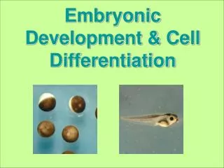
Differentiation and development. Development involves: 1) Cell division and differentiation Differentiation is the creation of different types of cells 2) Changes in anatomical structures Anatomical changes include g radual modification of physical and physiological characteristics

E N D
Differentiation and development • Development involves: • 1) Cell division and differentiation • Differentiation is the creation of different types of cells • 2) Changes in anatomical structures • Anatomical changes include gradual modification of physical and physiological characteristics • Development begins at fertilization
Stages of development • Development can be divided into: Prenatal and postnatal development • Prenatal development begins at fertilization and ends with birth • Prenatal development includes: • Embryological development • Changes occurring during the first two months after fertilization • Fetal development • Begins at the start of the ninth week and continues until birth • Postnatal development • Commences at birth and continues to maturity
Fertilization (conception) • Fertilization is fusion of two haploid gametes (egg and sperm) each with 23 chromosome to produce a zygote that contains 46 chromosomes • Fertilization occurs in the uterine tubes • Within a day of ovulation • Spermatozoa cannot fertilize an ovum until after capacitation
Fertilization Steps • Step 1 Ovulation • At ovulation the oocyte is in metaphase of meiosis II • Both the occyte and the polar body is surrounded by corona radiate • Step 2 Fertilization • Oocyte is surrounded by the sperms • Acrosomal enzyme from several sperms creates gaps in corona radiata • One sperm makes contact with the oocyte membrane • Sperm and oocyte fused • The process of meiosis will complete
The First Trimester • The first trimester is the period of embryological and early fetal development • Four processes occur during the first trimeter • 1) Cleavage • 2) Implantation • 3) Placentation • 4) Embryogenesis
The First Trimester • Cleavage - cleavage is the first cell division • The first cleavage occurs at 32 hours • Second cleavage occurs at 60 hour • Third cleavage occurs at 72 hours • Several divisions later the embryo becomes a solid mass of cells known as morula • Molurla becomes a blastocyst (also known as blastula) • Blastocyte includes: • Trophoblast – outer layer of cells that give rise to the chorion and placenta • Inner cell mass – cluster of cells at one end of blastocyst • Blastocoel – hallow fluid-filled inner cavity Cleavage
The First Trimester Implantation and Placentation • Implantation • Attachment of blastocyst into the uterine endometrium • The uterus is prepared for implantation by the hormone progesterone (see reproductive and endocrine lectures) • Occurs about 7 days after fertilization • Placentation • Blood vessels form around blastocyst and placenta develops • The placenta is a complex organ that permits exchange between the maternal and embryonic circulatory systems
The First Trimester • Gastrulation • After implantation, cell migration transforms blastula (single cell layer) to gastrula (3 layered structure) • Gastrulation is the generation of 3 distinct germ (cell) layers • Endoderm • Mesoderm • Ectoderm
Germ layers • Gastrulation • By day 12 surface cells move toward the primitive streak • A third germ layer forms • The three germ layers are: • Ectoderm – superficial cells that did not migrate • Will become integument (epidermis, hair, nail, epithelium, nose, mouth and anal canal), lense of eye, ear and nervous system • Endoderm – cells facing the blastocoele • Will become liver, pancreas, thyroid, bladder, lungs, epithelium of digestive and respiratory systems. • Mesoderm – migrating cells between ectoderm and endoderm • Will become musculskeletal system, circulatory system, excretory system, gonads, connective tissue of digestive and respiratory systems, smooth and cardiac muscle.
Germ layers • Organs and glands are consist of or include more than one germ layer • Adrenal cortex is derived from mesoderm • Adrenal medulla is derived from ectoderm
Neurulation • By the end of gastrulation, regions of the germ layers begin to develop into a rudimentary nervous system • Neurulation is the process of rudimentary nervous system formation • Notochord – a rod of mesodermal cells that develop along the axis. • Notochord cause ectoderm to bend inward and form a groove • Ectoderms grow on either side of the groove and then fuse together to form a neural tube • Neural tube becomes central nervous system (Brain and spinal cord)
Fetal Respiration • Growing fetus receives oxygen and eliminates carbon dioxide through a specialized circulatory system • The two components of this system are: • Placenta • Umbilical cord • Placenta and umbilical cord are outgrowths of the 4 extra-embryonic membrane (Amnion, Chorion, Allantois and yolk sac) that are formed during the development • Chorion extends villi into uterine wall and these villi become closely associated with endometrial cells • Chorionic villi forms intricate branching network for maternal blood • Umbilical cord connects fetus to placenta
Extraembryonic Membranes • Four extraembryonic membranes: • Yolk sac • Amnion • Allantois • Chorion
Embryo Anatomy • Yolk sac • Important site of blood cell formation • Amnion • Encloses fluid that surrounds and cushions developing embryo • Allantois • Eventually becomes bladder • Chorion • Placenta formation begins for chorion • Chorion is a membrane that completely surrounds amnion
Hormones of the placenta • Trophoblast secretes hormones to maintain pregnancy • Human chorionic gonadotropin (hCG) – Prevents disintegration of corpus luteum • At the ovaries corpus luteum produce progesterone • Estrogens – in addition to having role in secondary sexual characteristics it is involved in the thickening of the endometrium • Progesterone - enriches the uterus with a thick lining of blood vessels and capillaries so that it can sustain the growing fetus • Human Placental Lactogen (hPL) - decreases maternal insulin sensitivity, and therefore raises maternal blood glucose levels, whilst decreasing maternal glucose utilization, which helps ensure adequate fetal nutrition • Placental prolactin- stimulates the mammary glands to produce milk • Increased serum concentrations of prolactin during pregnancy cause enlargement of the mammary glands of the breasts and increases the production of milk. However, the high levels of progesterone during pregnancy stop ejection of milk. • After childbirth progesterone decreases and milk ejection initiates. • Relaxin
Second and Third Trimesters • Second trimester • Organ systems increase in complexity • Third trimester • Many organ systems become fully functional • Fetus undergoes largest weight change • At end of gestation fetus and uterus push maternal organs out of position
Developing fetus totally dependent on maternal organs • Maternal adaptations include increased • Respiratory rate • Tidal volume • Blood volume • Nutrient and vitamin uptake • Glomerular filtration rate
Structural and Functional Changes in the Uterus • Progesterone inhibits uterine muscle contraction • Opposed by estrogens, oxytocin and prostaglandins • Multiple factors interact to produce labor contractions in uterine wall
Labor and Delivery • Stages of labor • Dilation • The cervix dilates and fetus moves toward cervical canal • Expulsion • The cervix completes dilation and fetus emerges • Placental • Ejection of the placenta Goal of labor is parturition