Download
1 / 47
470 likes | 590 Views
Health Assessment Mary Ann Hudson, RN The Ohio State University College of Nursing. Nose. Ears. Mouth. Throat. Why does the RN assess the ears?. congenital ( syphilis + other TORCH infxns ) nerve damage CN VIII infant ear infections ( bottle-fed and Infant anatomy )

E N D
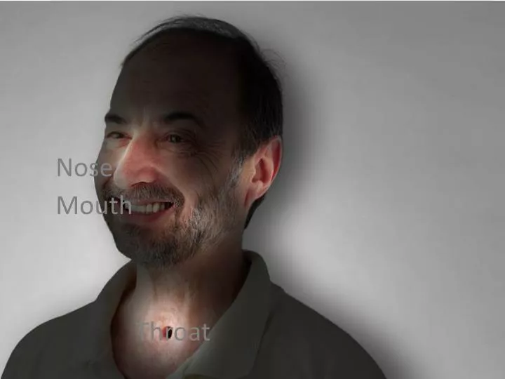
Health AssessmentMary Ann Hudson, RNThe Ohio State University College of Nursing Nose Ears Mouth Throat
Why does the RN assess the ears? • congenital (syphilis + other TORCH infxns) • nerve damage CN VIII • infant ear infections (bottle-fed and Infant anatomy) • structural changes (wax, trauma, infection) • effects from medications (tinnitus, vertigo)
Changes in the Elderly • Presbycusis Prez bĭ cū’ sĭs Sensorineural loss CN VIII Loss of high pitched sounds> 50 y.o. • Whisper • Consonants • Vowels http://www.youtube.com/watch?v=4YWSerwlWjM&feature=related
Changes in the Elderly • Otosclerosis of the middle ear • 20 – 40 y.o. • Hardening of stapes • Hearing in general is decreased gradually
Why does the RN assess the nose? To detect: • congenital anomalies • effects from trauma / obstruction • effects of illness (cancer, infections, allergies, nerve damage)
Changes in the Elderly • Smell diminishes slightly (anosmia) What are the implications to ADLs?
Why does the RN assess the mouth & throat? • congenital anomalies • tooth decay (bottles, hygiene) • nerve damage (swallow, gag) • effects of illness (cancer, infections, neurological & systemic disorders)
Changes in the Elderly • Xerostomia, less saliva & effects of meds • Receding gums; tooth loss • Taste ↓’s 80% (effect on ADLs?) • More lesions
Questions for the ENMT Interview Chief complaint: • Sensory perception, dryness, drainage, itching, pain, lesions Past History: • Injuries and accidents • Infections, allergies • Chronic illnesses, meds Social • Tobacco, Alcohol, Drugs/Rx and Abuse Environment • Noise, sun fumes, dust
Physical exam of the external structures of the ear Inspect • position, color, size and lesions Palpate • tenderness (auricle and tragus)
External Otitis Right 4x2 centimeter lesion involving tragus, outer canal, and lobule. Lesion significant for swelling, erythema, and purulent discharge. Tragus and pinna positive for pain 7/10 on palpation.
Otitis Media Assessment • Otoscopic examination Describe • Erythema, absent light reflex, swelling or bulging, visible bony landmarks, color, devices (tympanostomy tube), discharge, bleeding. Etiology • Supine bottle feed • 2nd hand smoke • Group daycare • Juvenile anatomy, genetics
Cauliflower Ear Etiology • Distortion of the cartilage due to relatively poor perfusion and venous drainage of pinna after blunt trauma. Can lead to obstructive hearing loss and may need surgical correction.
Tophi • Deposits of sodium biurate in the helix of the ear in uncontrolled states of gouty arthritis. May or may not be painful on palpation. Rule out other lesion etiology via history.
Mastoid air cells are open, air-containing spaces in one of the skull bones.
Mastoiditis Inspection is incredibly important! • infection of the bony air cells in the mastoid bone • drainage from the ear and redness (erythema) behind the ear over the mastoid bone • Forward displacement displacement of pinna • Abscess over mastoid process is possible. Tenderness, erythema, swelling and warmth appear over mastoid process. • Trace around border of erythema with felt-tip pen and document time of tracing.
Inspection of the internal structures Cerumen • Ear canal rich with specialized goblet cells that secrete cerumen which protects the ear canal with antibacterial and mechanical measures. • Cerumen varies by ethnicity and genetics. • Q-tips can damage or pack cerumen. • Ceremun impaction may create conductive hearing losses, pain or irritation and are most often a result of physical insertion of objects as cerumen will collect naturally at entrance of canal and can be wiped away as a part of normal hygiene. Commercial ear wax removers or provider removal is rarely needed.
Inspection of the internal structures Otitis Externa/Swimmer’s Ear • Moisture expands the cerumen and tissues of canal providing matrix for opportunistic bacteria to overgrow. May also arise from foreign body. • Erythema, tragus pain, exudate, and obstruction of canal due to swelling are typical presentation. • Patient may need to have wick inserted for otic antibiotic drops to reach site due to swelling.
Inspection of the internal structures Normal tympanic anatomy • Cone of light is situated toward the face • Bony landmarks should be visualized • “Pearly gray” membrane, but wide range of colors. Compare one side to the other. • Describe your findings: color, side to side differences, lesion/erythema location (use the face of the clock), exudate (in front of or behind TM), devices, cerumen disrupting view, scars (appear as bright white lesions), fluid behind TM (appear as bubbles behind TM).
Otoscopic assessment • Client tips head away from examiner. With infants and young children, have caregiver secure head (against caregiver shoulder, for example). • Use correctly sized speculum. Insert otoscope into auditory meatus. • Pull pinna of ear up and back for adults, down for children. Canal is a flexible invagination into bony structures of skull, use pinna and speculum to “drive” canal until TM can be visualized clearly. • Speculum will clear normal amounts of ceremun away for sufficient visualization of TM.
Otitis Media • Inflammatory involvement of TM and structures behind the TM within middle and inner ear. • Most often will see systemic signs and symptoms (fever, upper respiratory). • Common in infants and children due to anatomical development of eustachian tubes. • Describe absence of cone of light, TM inflammation and erythema, exudate, rupture (usually at margin of TM where it meets the canal, but can be anywhere on TM), “bulging” of TM outward from middle ear, loss of visualization of bony landmarks, marked difference between ears. • Current guidelines require 6 AOM in 1 year to be evaluated for TM tubes. • May lead to effusions (fluid behind TMs), or other anatomical changes like scarring, lesions, changes in bone morphology.
How to irrigate • Mineral oil and H2O2 (OTC preparations are Vibrox, Cerumen-X, and Murine). • Warm H2O with bulb syringe or water pik (cold water may cause syncope, vertigo, or nausea. Hot water will damage canal and set up patient for otitis externa). • Direct toward posterior wall of canal (not TM). • Don’t irrigate with tympanic perforation. • Patient should get up and walk slowly, with RN support as irrigation may disrupt balance dramatically. • Should not be a routine procedure.
Pediatric otoscopic exam • Do this system last. • Let child play with “flashlight.” • Important to hold still, may need restraint from caregiver. • Talking while examining is very loud for the child. • Hold handle of otoscope downwards as the curve of the relatively large infant/child head will limit angle of otoscope help with handle up (against head). • Use correctly sized speculum.
Hearing Screens Weber Test (CN VIII) • Tuning fork on middle forehead or top of head • Negative test is normal (i.e. no lateralization—positive is abnormal and is lateralization to either a “good” or “bad” side) • Bypass the external and middle ear
Hearing Screens Weber Test • Sound “lateralizes” to the affected (bad) ear (is heard loudest in bad ear because pitch from fork bypasses ear structures and skull transmits sound directly to sensory nerves in inner ear. Lack of hearing through the meatus is what makes the ear “bad” and the skull vibration louder in “bad” ear). • impacted cerumen • perforated ear drum • middle ear infection • foreign body • otosclerosis
Hearing Screens Weber Test • Sound “lateralizes” to the unaffected (good) ear. If inner ear sensory motor nerves cannot transmit sound to brain, tuning fork pitch will only be heard by the good ear. • Acoustic nerve damage (drugs, loud noises, brain insult). • Congenital
Hearing Screens Rinne test (CN VIII): • Tuning fork on mastoid process begin timing. • When patient raises hand to note that has ringing stopped (time ringing from placement on MP to a raised hand), move tines to external meatus and begin timing, patient raises hand when ringing stops again. • Normal is Air Conduction >Bone Conduction in both ears (A.U.)
Hearing Screens Rinne Test • If BC is longer or equal, there is a conduction loss • If there is no BC or AC, there is a sensorineural loss
Hearing Screens Voice - Whisper (CN VIII): • Client occludes one ear • Nurse whispers 2-3 syllables from 2 feet behind client (how are you?) • Normal=50% accuracy • Normal conversational speech is 40 decibels.
Physical exam of the nose Patency • Client occludes one nare while breathing through nose. • Infants are obligate nose breathers. If they cannot feed, their nares are not patent. • Assess for lesions, obstruction, and discharge by patient tipping head back, pulling tip of nose towards bridge, and using a light source into the nares. Internal structures • Turbinates • Septum • Mucous membranes • Describe lesions, color, anatomy visualized. When would a physical exam of the nose be particularly important?
Physical exam of the sinuses Frontal: • Deeply palpate at eyebrow level • Observe for periorbital swelling,symmetry, warmth, redness, nasal or lacrimal discharge, abscess. • Children to do not develop front sinus until puberty. Palpate ethmoid sinus in between eyebrows instead. Maxillary: • Deeply palpate medial to cheeks • Observe for mid-face swelling, symmetry, warmth, redness, nasal discharge, mouth pain, abscess (look into mouth).
Cranial Nerve I - Olfactory • Offer various scents for client to identify • Client occludes one narewhile smelling scent • Use scent readily available and easy to identify (alcohol pad, toothpaste on gauze, coffee from tray).
Epistaxis Acute Treatment • Sit up • Tilt head forward • Pinch nose 5 – 15 min. Etiology • Dry mucous membranes. • Trauma • Lesion • Allergy
Physical Exam of the Mouth and Throat External Structures Inspect: • Breath: malodorous => systemic disease, poor oral hygiene • Lips: hydration, lesions, symmetry, injury, skin, color Palpate: • Lips: lesions • Perioral area: lesions, swelling, warmth, symmetry
Physical Exam of the Mouth and Throat Internal Structures (Oropharynx) Inspect: • Tongue, buccal mucosa, gums, palates, tonsils, uvula, teeth • Describe color, symmetry, lesions, exudate, anatomical variations, dentation, swelling, bleeding • Use penlight or scope light. Use tongue blade or gauze pad to hold tongue. Wear gloves. Palpate (with gloves): • Buccalmucosa and gums for lesions or tooth eruption (infants and children). Palpate palate for symmetry and cleft (infants).
Physical Exam of the Mouth and Throat Grade Tonsils if Present • 1+ normal • 2+ half way btw pillars and uvula • 3+ touching uvula • 4+ touching each other • Note presence of exudate, stones, and describe color. Note if size is bi or uni lateral
Abnormal Findings Leukoplakia • Response to long term irritation and inflammation of muscosal membranes. • Overwhelming majority are benign. • Document location, size, and patient history of lesion. • Also found in female genital muscosal tissue due to long term irritation.
Abnormal Findings Candidiasis/Oral Thrush • Opportunistic fungal infection secondary to changes in normal flora. Normal flora is protective and can be disrupted by infection (bacterial or viral), diet, stress, medication, lifestyle, or environment/habits. • Painful, thick, white plague over tongue and mucosal surfaces that cannot be scraped off (leukoplakia is painless). • Presents in “satellite pattern” with small plaques radiating from larger central plaques.
Abnormal Findings Xerostomia • Cracked, dry, reddened tongue indicative of severe dehydration. May also be caused by medication. • Other signs of dehydration will be present including chapped lips and tacky buccal mucosa. • Elderly and pediatric population vulnerable.
Abnormal Findings Smooth Tongue • Vitamin and mineral deficiencies including B vitamins and iron (anemia) • Smooth burning tongue • History should corroborate finding (alcoholism, Celiac’s, poor nutrition, cystic fibrosis).
Abnormal Findings Chancre • syphilitic lesion (painless). It is typically found periorally or on lips. It is often ulcerated and can be large, but it is painless and remits on its own. Canker Sore • inside the mouth on the mucosa. It will be painful, ulcerated, sensitive to heat, cold, and acid. The patient may have other GI complaints. Canker sores often recur. Fever blister or cold sore • Caused by the Herpes Simplex Virus. It is typically found periorally or on the lips. It is a painful and gradually ulcerated lesion that begins invisibly as the sensation of tingling. The first instance of HSV infection may include fever, facial swelling, flu-like symptoms. HSV is chronic and lesions recur, though the use of ant-viral medications may reduce occurrence or prevent it, especially if used after first occurrence.
Abnormal Findings Bruxsim • Chronic teeth grinding wears down surfaces of the teeth, making them vulnerable to decay and breakage. Can be caused by pathophysiologic states (like mandibular joint diseases), medications, lifestyle, stress, or patients may grind their teeth while sleeping and be unaware. Dentists can fit patients with appliances to prevent tooth wear.
Abnormal Findings Hyperplasia • Overgrowth of gum tissue that may need surgical intervention • Dilantin • Pregnancy • Puberty • Leukemia
Abnormal Findings Gingivitis • A bacterially mediated process that initiates an inflammatory process of the gingiva, causing loss of the gingiva tissues. • Documented and rated according to level of recession of gums away from insertion areas of teeth. • Interventions include mechanical removal of bacterial plaques (flossing, scraping), antimicrobials, and regular dental hygiene.
Physical Exam of the Cranial Nerves Cranial Nerve XII • stick out tongue • side-to-side • strength • “la-la-la” • deviation to one side or weakness is abnormal Palates and tonsils, CNs IX and X • Inspect palates for shape, color and lesions • Elevation of uvula when tongue blade is on middle third of tongue and patient says “ah” is normal. • Gag patient with blade on posterior 1/3 of tongue (very small percentage of population do not have gag reflex). Patient can swallow water instead of gagging.