Download
1 / 55
550 likes | 553 Views
Translation. Chemistry 256. Historical notes. “ Adapter ” molecules (tRNA) bring amino acids to mRNA – Crick (1955) Synthesis of polyribonucleic acids using polynucleotide phosproylase – Heppel, Ortiz and Ochoa (1957) Genetic code – Nirenberg and Matthaei (1961)

E N D
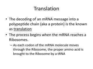
Translation Chemistry 256
Historical notes • “Adapter” molecules (tRNA) bring amino acids to mRNA – Crick (1955) • Synthesis of polyribonucleic acids using polynucleotide phosproylase – Heppel, Ortiz and Ochoa (1957) • Genetic code – Nirenberg and Matthaei (1961) • Insertion (or deletion) of triplet sequences of nucleotides causes minimal changes in expressed proteins – Crick, Brenner, Barnett and Watts-Tobin (1961) • First tRNA molecule sequence – Holley (1965)
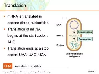
Genetic code • 20 amino acids need at least 20 different codes. • With four nucleotides, the smallest number needed to code for those amino acids is three (43 = 64 > 20). • There is a fair amount of redundancy built into the third position. For instance, serine is specified by the first two positions – UCX.
Reading frames determine message • Since codons are parsed in groups of three, the coded protein sequence depends on which nucleotide you start. • “Reading frame” indicates where the codons start, which is why insertion and deletion mutations are so serious. • An open reading frame contains no stop codons.
Structure of tRNA • Seemingly non-critical modifications (25% of bases are post-transcriptionally modified) may aid amino acid recognition. • Dihydrouridine (D) arm • Pseudouridine (ψ) arm • 15 invariant nt • 3’-CCA sequence is the amino acid acceptor • Variable arm is 3 – 21 nt long. • Anticodon loop
What holds it together? • Mostly stacking interactions, some base-pairing in the helical stems.
Aminoacyl-tRNA synthetase • Catalyzes the reaction to generate the aminoacyl-tRNA. • aaRS = amino acid-specific amino acyl tRNA synthetase. • Two classes of aaRS: class I does aminoacylation after anticodon recognition, class II does not even interact with anticodon.
So note the problem… • Given the number of bonds to be made, where is the power source? • Solution: Add ATP to amino acid and activate it; resulting mixed anhydride hydrolyzes a PPi and thus provides the energy needed.
Another two problems… • Enzyme must select the correct amino acid to attach to the tRNA; some of these enzymes don’t even recognize the anticodon. How do you prevent errors? • Due to the redundancy in the genetic code, there are only 20 tRNA molecules needed (one for each amino acid) and 64 anticodons. How do you make a particular tRNA “understand” that it might have to recognize up to four different anticodons?
Solution to the recognition problem: don’t even bother with the anticodon • Shown below is the yeast AspRS•tRNAAsp complex; it’s a dimer of the protein and there are two tRNAs bound to it. The key structural feature is the G30•U40 base pair in the anticodon stem (thus, not the anticodon). Due to this nonstandard pairing, this tRNA molecule can “bend” at this point whereas other tRNAs cannot, which allows only this tRNA to have Asp attached to it.
The fidelity of translation comes at the aminoacylation step, not at the ribosome • tRNA synthetase has a proofreading function, at a cost of an ATP per wrong aminoacylation. Mechanism is similar to proofreading in DNAP. • Yarus and Berg, “Phenylalanyl-tRNA Synthetase and Isoleucyl-tRNAPhe: A Possible Verification Mechanism for Aminoacyl-tRNA”, PNAS (1972) or Schreier and Schimmel “Transfer ribonucleic acid synthetase catalyzed deacylation of aminoacyl transfer ribonucleic acid in the absence of adenosine monophosphate and pyrophosphate”, Biochemistry (1972)
One other solution: many tRNAs anticodons bind to more than one codon • For instance, in the yeast tRNAPhe, the third base in the anticodon is methylated (2’-methyl guanosine) which allows it to not only hydrogen bond to C (normal) but to U as well. • Gm (2’-methyl guanosine) and I (inosine) are often found at the third position of the anticodon for amino acids with multiple redundancies. I binds to U, C and A.
“Wobble” hypothesis (Crick) explains the third position pairing ambiguity
Actually, there are more than 20 different tRNA molecules • In fact, there are at least 31 needed (using the wobble hypothesis base pairings). • In mammalian cells, there are in fact more than 150 different tRNAs, some of which attach to nonstandard amino acids, like selenocysteine (Sec).
Ribosomes – organelle where protein synthesis occurs (Zamecnik, 1955) Complex: made of both RNA strands and several proteins Size: big; 2500 kD in bacteria to 4500 kK in eukaryotes
Ribosome functions • Must bind mRNA with enough fidelity to recognize individual codons. • Must bind different tRNAs when needed. • Must interact with nonribosomal proteins that initiate, elongate and terminate protein synthesis. • Must catalyze peptide bond formation. • Must move so that sequential codons are translated.
Prokaryotic ribosomes • Two “units” to each ribosome; all are traditionally described in terms of sedimentation units. • Large and small units each comprise multiple RNA strands and multiple proteins. • 20,000 ribosomes in an E. coli cell makes up 80% of cell’s RNA content and 10% of its protein content.
Ribosomal RNA structure is defined by domains • Harry Noller (UC Santa Cruz, 1988) published the sequence of the 16S RNA of the small ribosomal subunit. • He expected stem loops to stabilize the structure, but the base pairing stems were short and imperfect.
X-ray crystallography revealed much more about the rRNA structure • Moore and Steitz (Yale, 2000) publish the structure, showing a lot of small irregular helical structures that fit into each other.
Distribution of protein and RNA in ribosomes is asymmetric • Proteins on both subunits tend to cluster in areas not associated with RNA (either mRNA or tRNA binding); the rRNA clusters where such binding occurs. • Most ribosomal proteins have a basic “tail” which binds to RNA using salt bridges.
Ribosomes bind three tRNAs, as well as mRNA • For all three sites, anticodon arms bind to 30S subunit and the rest of the tRNA binds to 50S subunit. • A (aminoacyl) site – binds incoming aminoacyl-tRNA • P (peptidyl) site – binds the growing peptide chain • E (exit) site – binds the deacylated tRNA
Peptidyl site details • The only one of the sites that will have a long molecule bound to it. • Growing peptide chain accommodated by a 100 Å-long tunnel in 50S subunit. • Lined with hydrophilic residues. • Barely large enough for an α-helix so no significant protein folding occurs at this stage.
Function of each subunit • Small subunit recognizes mRNA and tRNA, and binds them. • Large subunit catalyzes formation of peptide bond.
Eukaryotic ribosomes • Bigger and more complex than prokaryotic ribosomes. • Base-pairing differences indicate that structure is conserved, not sequence.
Peptide synthesis proceeds from N to C-terminus • Howard M. Dintzis (MIT), “Assembly of the peptide chains of hemoglobin”, PNAS (1961) showed tritium-labeled leucine being incorporated in the C-terminus of a growing hemoglobin chain.
Basic mechanism to make the peptide bond is an SN2 displacement • Note which of the tRNA binding sites these molecules are occupying. • The uncharged tRNA moves to the E site.
The work of ribosomes • Ribosomes read mRNA in the 5’ 3’ direction, which means that prokaryotes can literally have a RNAP make a transcript of the DNA and that transcript is immediately fed into a ribosome (can’t happen in eukaryotes due to transport through nuclear membrane). • Polysomes are multiple ribosome structures that bind to a single mRNA transcript.
Translation is intiated by fMet • Specifically, N-formylated methionine. • tRNAfMet differs from tRNAmet. • In prokaryotes, the same enzyme acylates the tRNAs; N-formylation occurs afterwards using another enzyme.
But how to set the reading frame (i.e., which AUG is really the “start”?) • Shine-Dalgarno sequence (Australia National Univ., 1974) is a purine-rich tract 10 nt upstream from initation codon. • This sequence is recognized and binds to the 16S ribosome subunit.
Pathway of mRNA through ribosome is a tunnel through 30S subunit • Note the A and P sites; the Shine-Dalgarno “anticodon” is located near the E site.
Initiation factors • Soluble proteins necessary to assemble the ribosome. • They drop off after the complex is assembled.
Initiation step 1 Initiation factor 3 helps dissociate the 50S subunit from the 30S, so that mRNA will have an easier time binding. IF-3 also assures that the A site is prepared to receive the fMet-tRNAfMet.
Initiation step 2 Shine, J. and Dalgarno, L. "Determinant of cistron specificity in bacterial ribosomes”, Nature (1975) 254, 34-38. The Shine-Dalgarno sequence, found in nearly all prokaryotic mRNAs, is complementary (at least in part) to the 16S rRNA unit in the small subunit; this helps situate the start codon correctly. IF-1 binds to the A site on the 30S subunit, preventing unintended tRNA binding.
Initiation step 3 GTP is finally hydrolyzed to rearrange the 30S subunit so that the fMet-tRNAfMet is in the P site, and IF-2 is removed from the complex. Eukaryotic initiation, though having similar steps, uses fundamentally different interactions. For instance, there are no Shine-Dalgarno sequences; eukaryotic mRNAs have a 7-methylguanosine cap and the first AUG encountered on the 5’ end is considered the start codon.
Elongation step 1 With the A site empty, the ribosome is ready to accept an amino acid-tRNA complex, which is also complexed to a GTP and the elongation factor Tu. The GTP hydrolyzes providing the energy to cause a conformational change in EF-Tu that “unlocks” the aa-tRNA to bind to the A site.
Elongation step 2 Transpeptidation occurs when the large subunit rRNA interacts with the tRNAs, the amino acid on the tRNA in the A site and the peptide chain on the tRNA in the P site. The rRNA has a catalytic function, albeit by arranging the components in the reaction rather than actively promoting bond formation or breakage.
Elongation step 3 In translocation, the P site tRNA, now without its amino acid, moves to the E site. The peptidyl-tRNA in the A site moves to the P site, leaving the A site open for the next round of elongation.
Codon-anticodon interactions are monitored to prevent errors First, second and third codon (purple)-anticodon (yellow) base pairs that form in the T. thermophilus 30S subunit; rRNA are the other-colored areas. Note that the third codon-anticodon pair doesn’t really have as strong an association with the rRNA – fits with the “wobble” hypothesis.
Allosteric interactions allow non-Crick-Watson base-pairing to be rejected T. thermophilus 30S subunit, with and without binding to tRNA and mRNA. The red bases are the nucleotides on the rRNA that undergo conformational changes. G530 switches from syn to anti conformation; if this does not occur, then the tRNA does not bind to this complex.
Ribosome’s active site lies entirely within an RNA domain • Thus, the catalytic transpepsidase activity is done entirely by RNA. • The proximity of the sites, especially the A and P sites, allows small conformational shifts to move tRNA from site to site.
Tetrahedral intermediate shows the SN2 mechanism of the acyl addition reaction The critical reaction which adds the A site amino acid to the P site polypeptide involves a nucleophilic attack of A-site amino acid amine (bottom) on the P-site carbonyl carbon (just above it). The enzyme forms hydrogen bonds to the substrate to stabilize it.
Elongation step 1 Unlike initiation, the steps of eukaryotic elongation resembles the steps of prokaryotic elongation. Both of the subunits have A, P and E conformations, so a state can be defined as the combined conformations of both subunits. T = EF Tu
Elongation step 2 GTP is hydrolyzed to move the amino-acid part of the A site tRNA fully into the large subunit A site. EF-Tu leaves at this point to be recharged with GTP.
Elongation step 4 Key point during translocation is that the acyl (amino acid) end of the tRNA, which is imbedded in the 50S subunit, shifts conformation first.
Elongation step 5 Then, subsequent GTP hydrolysis compels the rest of the tRNA molecule to move to the new conformation. This assures no “backsies”.
Antibiotics bind and block protein synthesis sites Due to the complexity of elongation, these steps are vulnerable to being blocked by competitor molecules. The similarity between puromycin and tyrosyl-tRNA allows puromycin to occupy the A site and attach to the peptide chain; its 3’ amide prevents further peptide chain elongation.
Termination step 1 • Need release factors (RF); for instance, RF-1 recognizes and binds to UAA codon. • Even though stop codons don’t attach to tRNAs, various RFs do recognize and bind to the codon itself. • True for both prokaryotes and eukaryotes.
Termination step 2 The normal transpeptidation step results in water being added to the peptide rather than another amino acid. The effect is to free the peptide chain from the ribosome.