Download
1 / 1
10 likes | 15 Views
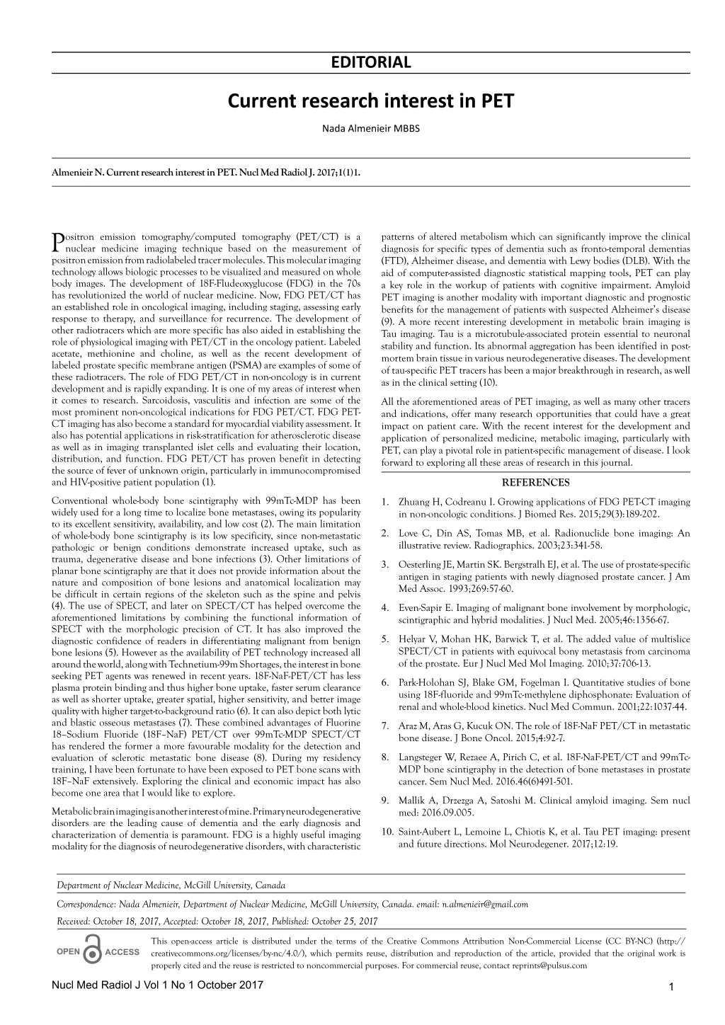
Positron emission tomography/computed tomography (PET/CT) is a nuclear medicine imaging technique based on the measurement of positron emission from radiolabeled tracer molecules. This molecular imaging technology allows biologic processes to be visualized and measured on whole<br>body images.

E N D
EDITORIAL Current research interest in PET Nada Almenieir MBBS Almenieir N. Current research interest in PET. Nucl Med Radiol J. 2017;1(1)1. P positron emission from radiolabeled tracer molecules. This molecular imaging technology allows biologic processes to be visualized and measured on whole body images. The development of 18F-Fludeoxyglucose (FDG) in the 70s has revolutionized the world of nuclear medicine. Now, FDG PET/CT has an established role in oncological imaging, including staging, assessing early response to therapy, and surveillance for recurrence. The development of other radiotracers which are more specific has also aided in establishing the role of physiological imaging with PET/CT in the oncology patient. Labeled acetate, methionine and choline, as well as the recent development of labeled prostate specific membrane antigen (PSMA) are examples of some of these radiotracers. The role of FDG PET/CT in non-oncology is in current development and is rapidly expanding. It is one of my areas of interest when it comes to research. Sarcoidosis, vasculitis and infection are some of the most prominent non-oncological indications for FDG PET/CT. FDG PET- CT imaging has also become a standard for myocardial viability assessment. It also has potential applications in risk-stratification for atherosclerotic disease as well as in imaging transplanted islet cells and evaluating their location, distribution, and function. FDG PET/CT has proven benefit in detecting the source of fever of unknown origin, particularly in immunocompromised and HIV-positive patient population (1). patterns of altered metabolism which can significantly improve the clinical diagnosis for specific types of dementia such as fronto-temporal dementias (FTD), Alzheimer disease, and dementia with Lewy bodies (DLB). With the aid of computer-assisted diagnostic statistical mapping tools, PET can play a key role in the workup of patients with cognitive impairment. Amyloid PET imaging is another modality with important diagnostic and prognostic benefits for the management of patients with suspected Alzheimer’s disease (9). A more recent interesting development in metabolic brain imaging is Tau imaging. Tau is a microtubule-associated protein essential to neuronal stability and function. Its abnormal aggregation has been identified in post- mortem brain tissue in various neurodegenerative diseases. The development of tau-specific PET tracers has been a major breakthrough in research, as well as in the clinical setting (10). ositron emission tomography/computed tomography (PET/CT) is a nuclear medicine imaging technique based on the measurement of All the aforementioned areas of PET imaging, as well as many other tracers and indications, offer many research opportunities that could have a great impact on patient care. With the recent interest for the development and application of personalized medicine, metabolic imaging, particularly with PET, can play a pivotal role in patient-specific management of disease. I look forward to exploring all these areas of research in this journal. REFERENCES Conventional whole-body bone scintigraphy with 99mTc-MDP has been widely used for a long time to localize bone metastases, owing its popularity to its excellent sensitivity, availability, and low cost (2). The main limitation of whole-body bone scintigraphy is its low specificity, since non-metastatic pathologic or benign conditions demonstrate increased uptake, such as trauma, degenerative disease and bone infections (3). Other limitations of planar bone scintigraphy are that it does not provide information about the nature and composition of bone lesions and anatomical localization may be difficult in certain regions of the skeleton such as the spine and pelvis (4). The use of SPECT, and later on SPECT/CT has helped overcome the aforementioned limitations by combining the functional information of SPECT with the morphologic precision of CT. It has also improved the diagnostic confidence of readers in differentiating malignant from benign bone lesions (5). However as the availability of PET technology increased all around the world, along with Technetium-99m Shortages, the interest in bone seeking PET agents was renewed in recent years. 18F-NaF-PET/CT has less plasma protein binding and thus higher bone uptake, faster serum clearance as well as shorter uptake, greater spatial, higher sensitivity, and better image quality with higher target-to-background ratio (6). It can also depict both lytic and blastic osseous metastases (7). These combined advantages of Fluorine 18–Sodium Fluoride (18F–NaF) PET/CT over 99mTc-MDP SPECT/CT has rendered the former a more favourable modality for the detection and evaluation of sclerotic metastatic bone disease (8). During my residency training, I have been fortunate to have been exposed to PET bone scans with 18F–NaF extensively. Exploring the clinical and economic impact has also become one area that I would like to explore. 1. Zhuang H, Codreanu I. Growing applications of FDG PET-CT imaging in non-oncologic conditions. J Biomed Res. 2015;29(3):189-202. 2. Love C, Din AS, Tomas MB, et al. Radionuclide bone imaging: An illustrative review. Radiographics. 2003;23:341-58. 3. Oesterling JE, Martin SK. Bergstralh EJ, et al. The use of prostate-specific antigen in staging patients with newly diagnosed prostate cancer. J Am Med Assoc. 1993;269:57-60. 4. Even-Sapir E. Imaging of malignant bone involvement by morphologic, scintigraphic and hybrid modalities. J Nucl Med. 2005;46:1356-67. 5. Helyar V, Mohan HK, Barwick T, et al. The added value of multislice SPECT/CT in patients with equivocal bony metastasis from carcinoma of the prostate. Eur J Nucl Med Mol Imaging. 2010;37:706-13. 6. Park-Holohan SJ, Blake GM, Fogelman I. Quantitative studies of bone using 18F-fluoride and 99mTc-methylene diphosphonate: Evaluation of renal and whole-blood kinetics. Nucl Med Commun. 2001;22:1037-44. 7. Araz M, Aras G, Kucuk ON. The role of 18F-NaF PET/CT in metastatic bone disease. J Bone Oncol. 2015;4:92-7. 8. Langsteger W, Rezaee A, Pirich C, et al. 18F-NaF-PET/CT and 99mTc- MDP bone scintigraphy in the detection of bone metastases in prostate cancer. Sem Nucl Med. 2016.46(6)491-501. 9. Mallik A, Drzezga A, Satoshi M. Clinical amyloid imaging. Sem nucl med: 2016.09.005. Metabolic brain imaging is another interest of mine. Primary neurodegenerative disorders are the leading cause of dementia and the early diagnosis and characterization of dementia is paramount. FDG is a highly useful imaging modality for the diagnosis of neurodegenerative disorders, with characteristic 10. Saint-Aubert L, Lemoine L, Chiotis K, et al. Tau PET imaging: present and future directions. Mol Neurodegener. 2017;12:19. Department of Nuclear Medicine, McGill University, Canada Correspondence: Nada Almenieir, Department of Nuclear Medicine, McGill University, Canada. email: n.almenieir@gmail.com Received: October 18, 2017, Accepted: October 18, 2017, Published: October 25, 2017 This open-access article is distributed under the terms of the Creative Commons Attribution Non-Commercial License (CC BY-NC) (http:// creativecommons.org/licenses/by-nc/4.0/), which permits reuse, distribution and reproduction of the article, provided that the original work is properly cited and the reuse is restricted to noncommercial purposes. For commercial reuse, contact reprints@pulsus.com OPEN ACCESS Nucl Med Radiol J Vol 1 No 1 October 2017 1


![Current Area of Research Interest [Include graphic]](https://cdn1.slideserve.com/3268815/slide1-dt.jpg)