Download
1 / 13
130 likes | 138 Views
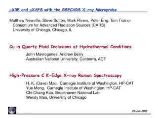
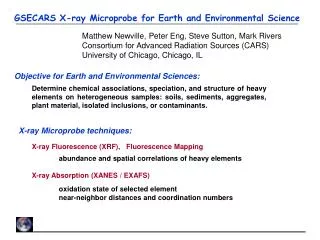
Matthew Newville, Steve Sutton, Mark Rivers, Peter Eng, Tom Trainor Consortium for Advanced Radiation Sources (CARS) University of Chicago, Chicago, IL. m XRF and m XAFS with the GSECARS X-ray Microprobe. Cu in Quartz Fluid Inclusions at Hydrothermal Conditions.

E N D
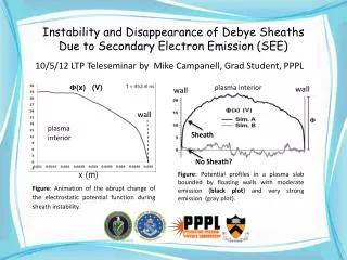
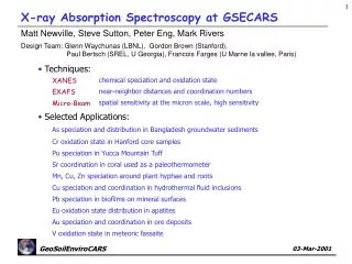
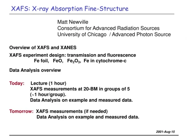
Matthew Newville, Steve Sutton, Mark Rivers, Peter Eng, Tom Trainor Consortium for Advanced Radiation Sources (CARS) University of Chicago, Chicago, IL mXRF and mXAFS with the GSECARS X-ray Microprobe Cu in Quartz Fluid Inclusions at Hydrothermal Conditions John Mavrogenes, Andrew Berry Australian National University, Canberra, ACT High-Pressure C K-Edge X-ray Raman Spectroscopy H. K. (Dave) Mao, Carnegie Institute of Washington, HP-CAT Yue Meng, Carnegie Institute of Washington, HP-CAT Chi-Chang Kao, Brookhaven National Lab Wendy Mao, University of Chicago
Advanced Photon Source Undulator A Period length 3.30 cm Number of periods 72 Length 2.47 m Minimum gap 10.5 mm Power (closed gap) 6 kW Kmax(closed gap) 2.78 Energy Tuning Range: 2.9 - 13.0 keV (1st harmonic) 2.9 - 45.0 keV (3rd and 5th harmonic) On-axis peak brilliance (at 6.5 keV): 9.6x10¹⁸ ph/s/mrad2 /mm2 /0.1%bw On-axis power density (closed gap): 167 kW/mrad2 Source Size and Divergence: Vert: s = 16mm, s’ = 4mrad Horiz: s = 240mm, s’= 14mrad
GSECARS Beamline Layout and Optics GeoSoilEnviroCARS: Sector 13, APS, Argonne National Lab Undulator Beamline:High collimation allows efficient focusing, for x-ray microprobe, and x -ray diffraction (small crystals, high pressure). High Pressure Station: Diamond-Anvil-Cell Large Volume Press X-ray Microprobe: XAFS, XRF, fluorescence tomography Monochromator: LN2-cooled Si (111) Energy range: 4.5 – 40keV Diffractometer: surface diffraction inelastic scattering Large Focusing Mirrors: 1m KB pair Storage Ring, undulator BM Station: tomography, diffraction, DAC, Large Volume Press, bulk XAFS Bending Magnet Beamline: 2nd-generation source, with high energy x-rays (up to 100KeV)
GSECARS XRF/XAFS Microprobe Station Focusing:Kirkpatrick-Baez mirrors: Rh-coated Si, typically using 3x3mm spot sizes, at 50mm from end of mirrors. Incident Beam: LN2 cooled Si (111) Sample Stage: x-y-z stage, 1mm resolution Slits:typically 200 to 300 mm, accepting ~20% of undulator beam at 50m from source. Data Collection: Flexible, custom software for X-Y XRF mapping, and XAFS, based on EPICS. Optical Microscope: 5x to 50x objective to external video system / webcam. Fluorescence detector:16-element Ge detector / DXP electronics, Lytle Detector, or Wavelength Dispersive Spectrometer
Kirkpatrick-Baez Focusing Mirrors The table-top Kirkpatrick-Baez mirrors use four-point benders and flat, trapezoidal mirrors to dynamically form an ellipsis. They can focus a 300x300mm beam to 1x1mm - a flux density gain of 105. With a typical working distance of 100mm, and an energy-independent focal distance and spot size, they are ideal for micro-XRF and micro-EXAFS. We use Rh-coated silicon for horizontal and vertical mirrors to routinely produce 2x3mm beams for XRF, XANES, and EXAFS.
16 element Ge Detector: energy resolution ~250 eV, which separates most fluorescence lines, and allow a full XRF spectrum (or the windowed signal from several lines) to be collected in seconds. Limited in total count rate (to ~250KHz), so multiple elements (10 to 30) are used in parallel. Detection limits are at the ppm level for XRF. XANES and EXAFS measurements of dilute species (~10ppm) in heterogeneous environments can be measured. X-ray Fluorescence Detectors Wavelength Dispersive Spectrometer has much better resolution (~20eV), and much smaller solid angle, but can be used for XAS, is able to separate fluorescence lines that overlap with a Ge detector.
Metal Speciation in Hydrothermal Fluid Inclusions 100mm John Mavrogenes, Andrew Berry (Australian National University) Hydrothermal ore deposits are important sources of Cu, Au, Ag, Pb, Zn, and U. Metal complexes in high-temperature, high-pressure solutions are transported until cooling, decompression, or chemical reaction cause precipitation and concentration in deposits. To further understand the formation of these deposits, the nature of the starting metal complexes need to be determined. XRF andXAFS are important spectroscopic tools for studying the chemical speciation and form of these metal complexes in solution. This is challenging to do at and above the critical point of water (22MPa, 375oC). Fluid inclusions from hydrothermal deposits can be re-heated and used as sample cells for high temperature spectroscopies. Natural Cu and Fe-rich brine / fluid inclusions in quartz from Cu ore deposits from New South Wales, Australia were examined at room temperature and elevated temperatures by XRF mapping and XAFS.
Hydrothermal Fluid Inclusion Measurements Linkham TS1500 Heating Stage. Normally, this can easily heat to 1200C for optical microscopy. We had to take off most of the protective front plates to cut down on background Cu and Fe fluorescence. In the end, we ran the quartz inclusion samples in air, with water flowing, but no heat shielding.
Cu speciation in Hydrothermal Fluid Inclusions Cu 25oC Fe 25oC Cu 495oC Fe 495oC 65mm XRF Mapping Understanding the metal complexes trapped in hydrothermal solutions in minerals is key to understanding the formation of ore deposits. Natural Cu and Fe-rich brine and vapor-phase fluid inclusions in quartz from Cu ore deposits were examined at room temperature and elevated temperatures by XRF mapping and EXAFS. Initial Expectation: chalcopyrite (CuFeS2) would be precipitated out of solution at low temperature, and would dissolve into solution at high temperature. We would study the dissolved solution at temperature. XRF mapping (2mm pixel size) showed that for large vapor-phase inclusions, a uniform distribution of Cu in solution at room temperature was becoming less uniform at temperature. This was reversible, and seen for multiple inclusions.
Cu XANES: Speciation in Fluid Inclusions XAFS measurements at low and high temperature for the vapor-phase inclusiong were also very different, with a very noticeable differences in the XANES: Low temp: Cu2+ , aqueous solution High temp: Cu1+ , Cl or S ligand. These results are consistent with Fulton et al [Chem Phys Lett. 330, p300 (2000)] study of Cu solutions near critical conditions: Cu2+ solution at low temperature, and Cu1+ associated with Cl at high temperatures.
Cu XAFS in Fluid Inclusions Cl O 2.35Å 2.09Å Cu2+ Cu1+ O 1.96Å EXAFS from the high temperature phase. Fit to high-temperature (450C) Cu solution in fluid (vapor phase) inclusion: can get good fits with 1 Cl at ~2.09Å and 1 O at ~2.00Å,or 2 Cl at ~2.08Å. This is also consistent with the model of for aqueous Cu1+ of Fulton et al, J. A. Mavrogenes, A. J. Berry, M. Newville, S. R. Sutton, Am. Mineralogist 87, p1360 (2002) Low temp High temp
- 6-element Si (440) Crystal Analyzer • - Kappa Diffractometer • Large Beamline KB mirrors, giving ~1013ph/s at 10keV in a 20x80mm spot. • Ideal for inelastic x-ray scattering in a Diamond Anvil Cell, including XANES-like information from X-ray Raman measurements. Inelastic X-ray Scattering: X-ray Raman DAC sample, lead-covered detector Analyzer Crystals: Si (440) 870mm Rowland circle
There is very little spectroscopic study of the phase transitions from graphite -> hcp C -> fcc C (diamond). Here is preliminary X-ray Raman measurements on graphite in a Diamond Anvil Cell (yes, background diamond is a possibility!) X-ray Raman: high pressure carbon