Download
1 / 25
280 likes | 676 Views
ANAEROBES. II BPT Dr Ekta Chourasia Department of Microbiology. Anaerobic Bacteria. Habitat : soil, water, decaying plants & animals. Intestines, mouth and the genital areas of animals and humans. Most of them are motile. Types of Anaerobic Bacteria.

E N D
ANAEROBES II BPT Dr Ekta Chourasia Department of Microbiology
Anaerobic Bacteria • Habitat : soil, water, decaying plants & animals. Intestines, mouth and the genital areas of animals and humans. • Most of them are motile. Dr Ekta, Microbiology, GMCA
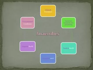
Types of Anaerobic Bacteria • Spore forming anaerobes : Only one genus- Clostridia - Gram-positive bacilli with round or oval spores which can be Terminal : Cl. tetani Sub-terminal : Cl. perfringens Central : Cl. bifermentans • Non-spore forming anaerobes – Many genus : both gram positive & gram negative bacteria. Cl. tetani Cl. perfringens Dr Ekta, Microbiology, GMCA
Leading Anaerobes CLOSTRIDIA • Cl. perfringens Gas gangrene • Cl. tetani Tetanus • Cl. botulinum Botulism (Food-poisoning) • Cl. difficile Pseudomembranous colitis Dr Ekta, Microbiology, GMCA
Cl. perfringens: Pathogenicities • Soft tissue infection: Gas gangrene (Type A) • Enteric infections: • Food poisoning (some strains of type A) • Gangrenous appendicitis (A + D) • Necrotising enteritis (C) • Biliary tract infections • Other diseases: • Brain abscess & meningitis • Thoracic infections Dr Ekta, Microbiology, GMCA
Gas Gangrene • Rapidly spreading, edematous myonecrosis which commonly involves wounds of muscles (battle wounds) that gets contaminated with spores of Cl. perfringens. • Spores can enter wounds with soil, road dusts, bits of clothing • Also called as malignant edema/ anaerobic myositis. • Toxic and invasive types i.e. it can spread along tissues • Many types of toxins are involved. The most important is lecithinase C / Alpha toxin Dr Ekta, Microbiology, GMCA
Clinical Presentation • Initially - no skin changes just pain • Systemic symptoms e.g. fever, dehydration • Once nerves damaged anaesthesia occurs • Paralysis • Skin changes - cellulitic progressing to dark purple; develop vesicles and bullae • Subcutaneous air on palpation (may not be present early on) • Foul smelling discharge • Oedema • Necrotic or haemorrhagic tissue • Patients may also present in septicaemic shock with tachycardia, hypotension, fever, stupor Dr Ekta, Microbiology, GMCA
Laboratory Diagnosis • Specimen – • films of muscles from the edge of infected area • exudates from the site of active infection • necrotic tissue & muscle fragment. • Microscopy – scanty pus cells, regularly shaped gram +ve bacilli with / without spores Dr Ekta, Microbiology, GMCA
Laboratory diagnosis • Culture Methods & Culture Media • McIntosh-Fildes’ jar method • Gaspak method • Anaerobic glove-box method • Robertson cooked meat medium (RCM) * Most of them are strict anaerobe but some can grow in the presence of less oxygen (aerotolerant) e.g. Cl.histolyticum Dr Ekta, Microbiology, GMCA
Laboratory Diagnosis • Stormy fermentation/ litmus milk reaction. • ‘Nagler reaction’- for the detection of Lecithinase C – seen as opalescence in serum or egg yolk containing media. Dr Ekta, Microbiology, GMCA
Treatment & Prophylaxis • Surgical debridement & wound irrigation. • Amputation – life threatening cases. • Hyperbaric oxygen • Antibiotics – Metronidazole • Passive immunization – anti gas gangrene serum. Dr Ekta, Microbiology, GMCA
Gas gangrene • Other causative agents : • Cl. septicum • Cl. novyi • Cl. histolyticum • Anaerobic streptococci • E.coli, proteus, staphylococci Dr Ekta, Microbiology, GMCA
Cl.tetani • Morphology : drum stick appearance non capsulated & motile • Culture : obligate anaerobe • Identification & toxigenicity testing – BA – hemolysis, Nagler reaction (tetanolysin) • Produces swarming • Produces 2 toxins: Tetanospasmin & Tetanolysin Dr Ekta, Microbiology, GMCA
Tetanus • Contamination of wound - surgical or accidental. • Injuries, septic abortions • Toxin mediated disease – locally produced Tetanospasmin - a neuro toxin : spread to the nerve endings and cause spasm of muscles. • Typical symptom - Lock-jaw : difficulty in opening mouth (known as Trismus). The severe muscle spasm end in respiratory failure. Dr Ekta, Microbiology, GMCA
Prophylaxis • Surgical prophylaxis – removal of foreign bodies, necrotic tissues • Antibiotics – to destroy or inhibit tetanus bacilli or other pyogenic bacteria in wounds • Immunisation Dr Ekta, Microbiology, GMCA
Prevention • Vaccination - Tetanus toxoid. • Children, pregnant women. • Tetanus neonatorum and uterine tetanus have high mortality rate. • Whenever cut or wounds occurs, vaccinate the individual with anti tetanus serum (ATS). Immunity last for 6-8 months only. Dr Ekta, Microbiology, GMCA
Treatment • In hospitals – isolated units • Protect them from noise & light • Maintain airway by tracheostomy • Human TIG – slow IV infusion • Penicillin or Metronidazole for a week or more • Full course of immunisation Dr Ekta, Microbiology, GMCA
Botulism • Most severe type of food-poisoning caused by Cl. botulinum. • Produces a powerful exotoxin - toxin binds to the neuron at N-M junction and prevents the release of acetylcholine across the synaptic cleft : Flaccid paralysis • By consuming foods contaminated with pre formed toxins - mainly meat and other products. • 3 types – food borne (preserved food), wound & infant • Death can occur due to respiratory failure Dr Ekta, Microbiology, GMCA
Botulism – treatment & prevention • Treatment is by anti-toxin and not by antibiotics. • All foods and mainly tinned foods should be cooked well before eating. The toxin is neutralised by cooking. • If the tinned food is giving bad smell or the tin is found bulging or gas comes out while opening the tin, do not consume such food. Dr Ekta, Microbiology, GMCA
Pseudomembranous Colitis • Often associated with antibiotics – also known as antibiotic associated colitis/ antibiotic associated diarrhoea. • Due to active multiplication of Cl. difficile& production of its toxins – enterotoxin & cytotoxin • Mild type of disease and can be treated by metronidazole. Vancomycin & bacitracin are also effective. • No vaccine is available. Dr Ekta, Microbiology, GMCA
Non - sporing Anaerobes Dr Ekta, Microbiology, GMCA
Habitat • Man, Animal and soil • In HUMANS: Intestine Mouth : Gingival crevice Vagina : Lactobacillus Skin : Propionibacterium (acne) Dr Ekta, Microbiology, GMCA
Brain abscesses Oro-pharyngeal infections Pleuro-pulmonary infections Intra-abdominal infections Abdominal abscesses Female genital tract infections Skin, soft tissue & bone infections Endocarditis Bacteremias Diseases caused by non sporing anaerobes Dr Ekta, Microbiology, GMCA