Download
1 / 30
310 likes | 976 Views
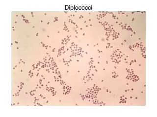
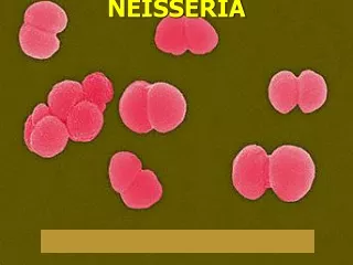
Neisseriae gram-negative cocci (diplococci) facultative anaerobes Neisseria gonorrhoeae - gonorrhea Neisseria meningitidis - meningitis and septicemia gonococci ( Neisseria gonorrhoeae ) obligate human pathogens sometimes carriers are asymptomatic

E N D
Neisseriae • gram-negative • cocci (diplococci) • facultative anaerobes • Neisseria gonorrhoeae - gonorrhea • Neisseria meningitidis - meningitis and septicemia • gonococci (Neisseria gonorrhoeae) • obligate human pathogens • sometimes carriers are asymptomatic • Gonorrhea - infection of epithelium of distal urethra or cervix • Ascent of bacteria - pelvic inflammatory disease (PID) • epididymitis
Gonococcal infection process I. Attachment of bacteria to columnar epithelial cells of distal urethra or cervix Pili and bacterial cell surface proteins II. Motility of ciliated cells slows/ceases III. Ciliated cells die and are sloughed off IV. Bacteria are internalized by nonciliated cells bacteria direct endocytosis V. Intracellular replication in phagocytic vacuoles VI. Intracellular transport to base of epithelial cells VII. Exocytosis - Fusion of vacuoles with plasma membrane release into connective tissue/local inflammation/dissemination
Damage caused by gonococcal infections due to LPS, peptidoglycan (induce inflammatory response) No exotoxin secreted by gonococci Gonococcal evasion of host immune response pili and bacterial cell surface proteins are immunodominant neutrophils can engulf gonococci BUT pili and bacterial cell surface proteins highly changeable neutrophils need bacterial cell surface proteins bacteria lacking Opa proteins are not engulfed
Strategies to evade the host immune response 1. Antigenic variation - changes in composition/structure of surface molecules not in response to antibodies due to genetic rearrangements (high frequency) reassortment and recombination of duplicated gene segments example: pili structure one complete pilin gene: pilE 10-15 variant pilin genes with 5’ truncations: pilS loci
Strategies to evade the host immune response 2. Phase variation - gene expression turned on/off A. gain/loss of repeat sequences at 5’ ends of genes -changes reading frame -due to DNA strand misalignment during replication (Opa) -strategy also used for LPS biosynthetic genes
Strategies to evade the host immune response 2. Phase variation - gene expression turned on/off B. site-specific inversion of gene promoter (fimA of E. coli)
Strategies to evade the host immune response 3. Production of IgA1 protease cleaves IgA1 in hinge 4. Modification of LPS with sialic acid -sialic acid a RBC cell surface component (camouflage) 5. Occupancy of intracellular environment
Meningococcal infections bacteria closely related to gonococci diseases different gonococcal - localized meningococcal - systemic and life threatening encounters usually asymptomatic (nasopharynx) disease mechanism cell penetration similar to gonococci differences: meningococci encapsulated and possess a hemolysin (exotoxin) in bloodstream - purpura fulminans (disseminated intravascular coagulation)
Diagnosis Gonococci: gram-negative diplococci in neutrophils from cervix/urethra Meningococci gram-negative diplococci in CSF or blood. culturing (chocolate agar) Treatment cephalosporin (b-lactamase resistant version) = ceftriaxone (IM)
Enteric bacteria Pathogenic varieties cause diarrheal diseases Watery diarrhea loss of electrolytes and fluids from small intestine severe cases can be fatal within hours (dehydration) Bloody diarrhea = dysentery local tissue damage or inflammation of intestinal tract Diarrheal diseases kill millions of children worldwide each year also result in malnutrition and growth retardation Diarrheal disease agents bacteria, viruses, and protozoa
Serotyping Classification method for E. coli (path. vs nonpath.) Lipopolysaccharides (LPS) • In outer membrane (gram-neg.) • consists of three parts • lipid A • core polysaccharide • O side chain (O antigen) • O antigen (LPS)(serogroup) • 173 different varieties • H antigen (flagellum)(serotype) • 60 different varieties K antigen (capsule) K1 antigen
Examples: E. coli O157:H7 is a pathogenic strain E. coli in O86 serogroup rarely pathogenic (normal flora) E. coli in O55 serogroup usually pathogenic (rarely in normal flora) Cholera bacteria also classified this way Vibrio cholera O1 (“classical” and “El Tor” strains) Vibrio cholera O139 Salmonella species over 2300 serotypes
Virotyping classification scheme based on virulence factors New method based on molecular properties Still undergoing progress as a system Particular virotype usually contains more than one serogroup or serotype Some E. coli strains have properties of more than one virotype Diffusely adhering (DAEC)
Where are these pathogens found? Vibrio cholera normally inhabits coastal waters humans infected from contaminated water or food Salmonella, E. coli, others not normally found in environment associated with humans and animals How are these organisms transmitted? Transfer is from feces to mouth (fecal-oral mode of transfer) 7 F’s: feces, food, fluids, fingers, flies, fomites, fornication Inoculum size needed for disease varies with organism Shigella dysenteriae - 100’s of cells ET E. coli - 108 cells
Why such a great difference in inoculum size? ETEC is not invasive must survive host defenses and g.i. tract environment pH ranges from 1 to 9 along tract mucus in small intestine (interfers with binding to host cells) moved along by peristalsis encounter soluble host proteins lysozyme proteases lipases IgA immunoglobulins bile salts host phagocytic cells resident bacteria take up available space produce bacteriocins (kill off competitors)
Adhere to epithelial cell surface adhesins = pili and or fimbriae Vibrio cholera toxin coregulated pilus (TcpA) = adhesin expression of TcpA is controlled by toxR gene toxR is like a switch (“master switch gene”) turned on by change from salt water to human intestine toxR also turns on gene for cholera toxin (CtxA) ctxA encoded on bacteriophage genome integrated in the genome of V. cholera
CtxA - ADP ribosylation of adenylate cyclase subunit (G protein subunit) ETEC LT (labile toxin) has same mechanism as CtxA ST (stable toxin) binds membrane guanylate cyclase, increases cGMP Similar-acting toxins produced by Salmonella, Campylobacter, Yersinia, Aeromonas, andEA E. coli
EPEC - disease not caused by a secreted toxin BfpA = bundle-forming pilus Signal transduction induced by binding phosphorylation of Hsp90 activation of phospholipase C increase calcium and IP3 Eventually rearrange cytoskeleton pedestal structure characteristic of EPEC
Diagnosis V. cholera often guess before definitive diagnosis E. coli O antigen test Treatment replace electrolytes and fluid loss (oral rehydration) usually self-limiting no vaccines available
The Shigellae S. dysenteriae serogroup A (distinguish by O antigen) S. flexneri serogroup B S. boydii serogroup C S. sonnei serogroup D Shigellae are differentiated pathogenic clones of E. coli Disease course very host specific disease course small inoculum (relatively acid resistant) Genes for acid resistance large intestine invade M cells
Invasion depends on OM proteins Bacteria released into lamina propria some ingested by macrophages some access basal surface of epithelial cells (gain entry into cells) IL-1 release induces inflammatory response neutrophils recruited migrate through mucosa allow bacteria through (other way) Bacteria escape phagosome Cell-to-cell invasion polymerize actin Neutrophils and blood in stool (symptomatic)
Shiga toxin (Stx) produced by some cells A-B toxin cleaves rRNA (stops protein synthesis) Diagnosis bloody diarrhea Leukocytes in stool culture Treatment oral rehydration antibiotics for severe cases (resistance problem) fluoroquinolones new beta-lactams and cephalosporins no vaccine
EHEC nonfebile bloody diarrhea (hemorrhagic colitis) E. coli O157:H7 acid resistant (small inoculum) Large intestine attach to gut mucosa (do not invade) attaching and effacing lesion on gut wall shigalike toxins - induce inflammation Complication - damage to small blood vessels at distant sites kidney - HUS (hemolytic uremic syndrome) acute renal failure (death)
Salmonella S. enterica gastroenteritis S. cholerasuis focal infection of vascular endothelium S. typhimurium focal infection of vascular endothelium infections of particular organ systems S. typhi, S. paratyphi Typhoid fever (human specific) Acid sensitive Penetrate mucosa barrier (microbe directed) Bacteremia not sustained Typhoid fever - multiply in macrophages bacteria shed continuously into bloodstream (septicemia) do have sustained bacteremia
Diagnosis - culture and O serotype Treatment - gastroenteritis, no antibiotics systemic, antibiotics