Download
1 / 1
10 likes | 127 Views
AP. A. B. A. B. LLat. C. D. C. D. Intra and Inter-Therapist Reproducibility of Daily Isocenter Verification Using Prostatic Fiducial Markers

E N D
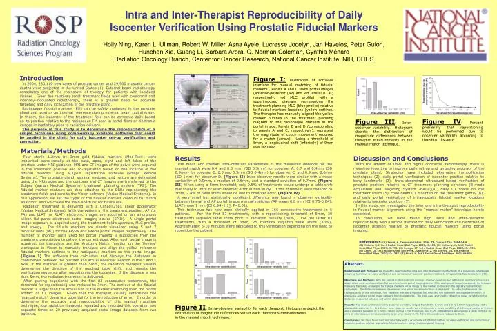
AP A B A B LLat C D C D Intra and Inter-Therapist Reproducibility of Daily Isocenter Verification Using Prostatic Fiducial Markers Holly Ning, Karen L. Ullman, Robert W. Miller, Asna Ayele, Lucresse Jocelyn, Jan Havelos, Peter Guion, Hunchen Xie, Guang Li, Barbara Arora, C. Norman Coleman, Cynthia Ménard Radiation Oncology Branch, Center for Cancer Research, National Cancer Institute, NIH, DHHS Introduction In 2004, 230,110 new cases of prostate cancer and 29,900 prostatic cancer deaths were projected in the United States (1). External beam radiotherapy constitutes one of the mainstays of therapy for patients with localized disease. Given the relatively small treatment fields used with conformal and intensity-modulated radiotherapy, there is a greater need for accurate targeting and daily localization of the prostate gland. Radiopaque fiducial markers (FM) can be safely implanted in the prostate gland and used as an internal reference during external beam radiotherapy. In theory, the isocenter of the treatment field can be corrected daily based on its position relative to the radiopaque FM seen in portal films or electronic images immediately prior to radiation delivery. The purpose of this study is to determine the reproducibility of a simple technique using commercially available software that could be applied in the clinic for daily isocenter set-up verification and correction. Figure I: Illustration of software interface for manual matching of fiducial markers. Panels A and C show portal images (anterior-posterior (AP) and left lateral (LLat) respectively, red MLC profile) with a superimposed diagram representing the treatment planning MLC (blue profile) relative to fiducial markers locations (yellow outline). The therapist has manually aligned the yellow marker outlines in the treatment planning diagram to the radiopaque markers in the portal image. Panels B and D (corresponding to panels A and C, respectively), represent the magnitude of couch movement required for a match (arrow). Using a threshold of 5mm, a longitudinal shift (inferiorly) of 9mm was required. Figure IV: Percent probability that repositioning would be performed due to observer variability according to threshold distance Figure III: Inter-observer variability. Histogram depicts the distribution of magnitude differences between therapist measurements in the manual match technique. Materials/Methods Four sterile 1.2mm by 3mm gold fiducial markers (Med-Tec) were implanted trans-rectally at the base, apex, right and left lobes of the prostate under MRI guidance. MRI and CT images of the pelvis were acquired in the treatment position and co-registered based on the location of the fiducial markers using ACQSIM registration software (Philips Medical Systems). The prostate gland, seminal vesicles, and rectum are delineated using the MRimages and the contours and CT images are transferred to the Eclipse (Varian Medical Systems) treatment planning system (TPS). The fiducial marker contours are then attached to the DRRs representing the treatment fields and sent to the Vision software (Varian Medical Systems). In this application, we set the ‘type’ of the fiducial markers contours to ‘match anatomy’, and we create the ‘field aperture’ for future use. Radiation treatment is delivered with a Clinac21EX linear accelerator (Varian Medical Systems). With the patient in the treatment position, AP (or PA) and LLAT (or RLAT) electronic images are acquired on an amorphous silicon flat panel electronic portal imaging device (EPID). A single portal image exposure is acquired using the treatment field’s aperture, MLC profile, and energy. The fiducial markers are clearly visualized using 5 and 7 monitor units (MU) for the AP/PA and lateral portal images respectively. The number of monitor units used for portal imaging is subtracted from the treatment prescription to deliver the correct dose. After each portal image is acquired, the therapists use the ‘Anatomy Match’ function on the ‘Review’ workspace in Vision to manually translate and align the yellow reference fiducial markers outlines to the radiopaque markers on the portal image. (Figure I) The software then calculates and displays the distances in centimeters between the planned and actual isocenter location in the Y and X axis. If the distance is greater than 5mm, the radiation therapist visually determines the direction of the required table shift, and repeats the verification sequence after repositioning the isocenter. If the distance is less than 5mm, the radiation treatment is delivered. After gaining experience with the first 83 consecutive treatments, the threshold for repositioning was reduced to 3mm. The contour of the fiducial marker is larger than the actual size of the marker stemming from the bloom artifact on CT images. Given that the therapist visually determines the ‘manual match’, there is a potential for the introduction of error. In order to determine the accuracy and reproducibility of this manual matching technique, four radiation therapists repeated and recorded this operation two separate times on 20 previously acquired portal image datasets from two patients. Results The mean and median intra-observer variabilities of the measured distance for the manual match were 0.4 and 0.3 mm (SD 0.5mm) for observer A, 0.7 and 0.4mm (SD 0.9mm) for observer B, 0.5 and 0.5mm (SD 0.4mm) for observer C, and 0.9 and 0.6mm (SD 1mm) for observer D. (Figure II) Inter-observer results were similar with a mean variability of 0.9mm, a median of 0.6mm, and a standard deviation of 0.7mm. (Figure III) When using a 5mm threshold, only 0.5% of treatments would undergo a table shift due solely to intra or inter-observer error in this study. If this threshold were reduced to 3mm, 2.4% of table shifts would be due to observer error. (Figure IV) A very small but statistically significant difference was found in observer variability between lateral and AP portal image manual matches (AP mean 0.8 mm [CI 0.75-0.84], LLAT mean 1 mm [CI 0.94–1.1], P<0.01). This technique has now been clinically applied in 166 consecutive treatments in 6 patients. For the first 83 treatments, with a repositioning threshold of 5mm, 30 treatments required table shifts prior to radiation delivery (36%). For the latter 83 treatments, with a threshold of 3mm, 25 fractions required table shifts (30%). Approximately 5-10 minutes were dedicated to this verification depending on the need to reposition the patient. Discussion and Conclusions With the advent of IMRT and highly conformal radiotherapy, there is mounting incentive to improve daily set-up and targeting accuracy of the prostate gland. Strategies have included alternative immobilization techniques (2), daily portal verification of isocenter position relative to bony landmarks (3), trans-abdominal ultrasound-based verification of prostate position relative to CT treatment planning contours (B-mode Acquisition and Targeting System -BAT)(4), daily CT scans on the treatment couch (5), cone-beam CT mounted on the treatment gantry (6), and daily portal verification of intraprostatic fiducial marker locations relative to isocenter position (7). In this study, we investigated the inter and intra-therapist reproducibility in fiducial marker alignment using the “manual match” technique herein described. In conclusion, we have found high intra and inter-therapist reproducibility with a simple method for daily verification and correction of isocenter position relative to prostatic fiducial markers using portal imaging. References: (1) Jemal, A, Cancer statistics, 2004. CA Cancer J Clin. 2004;54:8. (2) Malone, S, J. Int J Radiat Oncol Biol Phys. 2000;48:105. (3) Hatherly, K, Int J Radiat Oncol Biol Phys. 1999;45:791. (4) Serago, CF, Int J Radiat Oncol Biol Phys. 2002;53:1130. (5) Court, LE, Int J Radiat Oncol Biol Phys. 2004;59:412. (6) Jaffray, DA, Int J Radiat Oncol Biol Phys. 2002;53:1337. (7) Alasti, H, Int J Radiat Oncol Biol Phys. 2001;49:869. Abstract: Background and Purpose: We sought to determine the intra and inter-therapist reproducibility of a previously established matching technique for daily verification and correction of isocenter position relative to intraprostatic fiducial markers (FM). Materials and Methods: With the patient in the treatment position, anterior-posterior and left lateral electronic images are acquired on an amorphous silicon flat panel electronic portal imaging device. After each portal image is acquired, the therapist manually translates and aligns the fiducial markers in the image to the marker contours on the digitally reconstructed radiograph. The distances between the planned and actual isocenter location is displayed. In order to determine the reproducibility of this technique, four radiation therapists repeated and recorded this operation two separate times on 20 previously acquired portal image datasets from two patients. The data were analyzed to obtain the mean variability in the distances measured between and within observers. Results: The mean and median intra-observer variability ranged from 0.4 to 0.7mm and 0.3 to 0.6mm respectively with a standard deviation of 0.4 to 1.0mm. Inter-observer results were similar with a mean variability of 0.9mm, a median of 0.6mm, and a standard deviation of 0.7mm. When using a 5 mm threshold, only 0.5% of treatments will undergo a table shift due to intra or inter-observer error, increasing to an error rate of 2.4% if this threshold were reduced to 3mm. Conclusion: We have found high reproducibility with a previously established method for daily verification and correction of isocenter position relative to prostatic fiducial markers using electronic portal imaging. Figure II: Intra-observer variability for each therapist. Histograms depict the distribution of magnitude differences within each therapist’s measurements in the manual match technique.