Download
1 / 9
160 likes | 468 Views
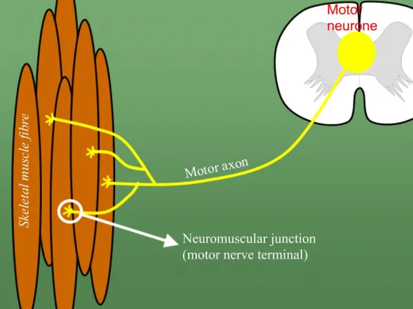
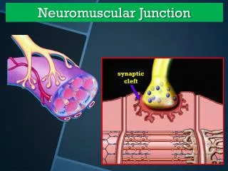
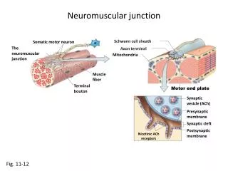
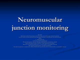
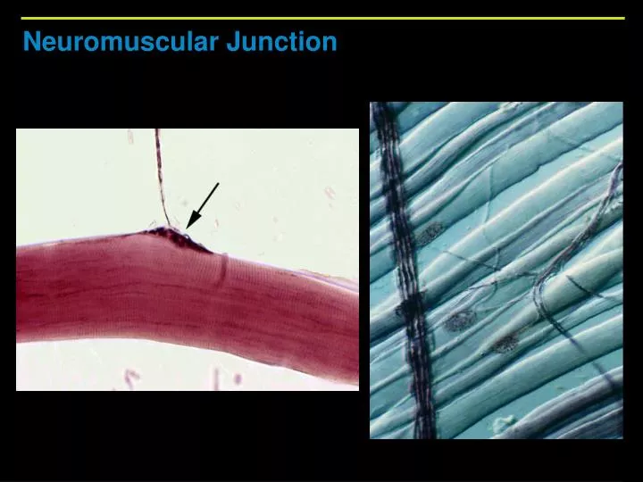
Neuromuscular Junction. Neurotransmitter released diffuses across the synaptic cleft and attaches to ACh receptors on the sarcolemma. Axon terminal. Synaptic cleft. Synaptic vesicle. Sarcolemma. T tubule. 1. Net entry of Na + Initiates an action potential which

E N D
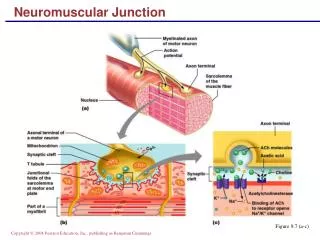
Neurotransmitter released diffuses across the synaptic cleft and attaches to ACh receptors on the sarcolemma. Axon terminal Synaptic cleft Synaptic vesicle Sarcolemma T tubule 1 Net entry of Na+ Initiates an action potential which is propagated along the sarcolemma and down the T tubules. ACh ACh ACh Ca2+ Ca2+ SR tubules (cut) SR Ca2+ Ca2+ 2 Action potential in T tubule activates voltage-sensitive receptors, which in turn trigger Ca2+ release from terminal cisternae of SR into cytosol. ADP Pi Ca2+ Ca2+ Ca2+ Ca2+ 6 Tropomyosin blockage restored, blocking myosin binding sites on actin; contraction ends and muscle fiber relaxes. 3 Calcium ions bind to troponin; troponin changes shape, removing the blocking action of tropomyosin; actin active sites exposed. Ca2+ 5 Removal of Ca2+ by active transport into the SR after the action potential ends. Ca2+ 4 Contraction; myosin heads alternately attach to actin and detach, pulling the actin filaments toward the center of the sarcomere; release of energy by ATP hydrolysis powers the cycling process. Figure 9.10
Neurotransmitter released diffuses across the synaptic cleft and attaches to ACh receptors on the sarcolemma. Axon terminal Synaptic cleft Synaptic vesicle ACh ACh ACh Figure 9.10
Neurotransmitter released diffuses across the synaptic cleft and attaches to ACh receptors on the sarcolemma. Axon terminal Synaptic cleft Synaptic vesicle Sarcolemma T tubule 1 Net entry of Na+ Initiates an action potential which is propagated along the sarcolemma and down the T tubules. ACh ACh ACh Figure 9.10
Neurotransmitter released diffuses across the synaptic cleft and attaches to ACh receptors on the sarcolemma. Axon terminal Synaptic cleft Synaptic vesicle Sarcolemma T tubule 1 Net entry of Na+ Initiates an action potential which is propagated along the sarcolemma and down the T tubules. ACh ACh ACh Ca2+ Ca2+ SR tubules (cut) SR Ca2+ Ca2+ 2 Action potential in T tubule activates voltage-sensitive receptors, which in turn trigger Ca2+ release from terminal cisternae of SR into cytosol. Ca2+ Ca2+ Ca2+ Ca2+ Figure 9.10
Neurotransmitter released diffuses across the synaptic cleft and attaches to ACh receptors on the sarcolemma. Axon terminal Synaptic cleft Synaptic vesicle Sarcolemma T tubule 1 Net entry of Na+ Initiates an action potential which is propagated along the sarcolemma and down the T tubules. ACh ACh ACh Ca2+ Ca2+ SR tubules (cut) SR Ca2+ Ca2+ 2 Action potential in T tubule activates voltage-sensitive receptors, which in turn trigger Ca2+ release from terminal cisternae of SR into cytosol. Ca2+ Ca2+ Ca2+ Ca2+ 3 Calcium ions bind to troponin; troponin changes shape, removing the blocking action of tropomyosin; actin active sites exposed. Ca2+ Figure 9.10
Neurotransmitter released diffuses across the synaptic cleft and attaches to ACh receptors on the sarcolemma. Axon terminal Synaptic cleft Synaptic vesicle Sarcolemma T tubule 1 Net entry of Na+ Initiates an action potential which is propagated along the sarcolemma and down the T tubules. ACh ACh ACh Ca2+ Ca2+ SR tubules (cut) SR Ca2+ Ca2+ 2 Action potential in T tubule activates voltage-sensitive receptors, which in turn trigger Ca2+ release from terminal cisternae of SR into cytosol. Ca2+ Ca2+ Ca2+ Ca2+ 3 Calcium ions bind to troponin; troponin changes shape, removing the blocking action of tropomyosin; actin active sites exposed. Ca2+ 4 Contraction; myosin heads alternately attach to actin and detach, pulling the actin filaments toward the center of the sarcomere; release of energy by ATP hydrolysis powers the cycling process. Figure 9.10
Neurotransmitter released diffuses across the synaptic cleft and attaches to ACh receptors on the sarcolemma. Axon terminal Synaptic cleft Synaptic vesicle Sarcolemma T tubule 1 Net entry of Na+ Initiates an action potential which is propagated along the sarcolemma and down the T tubules. ACh ACh ACh Ca2+ Ca2+ SR tubules (cut) SR Ca2+ Ca2+ 2 Action potential in T tubule activates voltage-sensitive receptors, which in turn trigger Ca2+ release from terminal cisternae of SR into cytosol. Ca2+ Ca2+ Ca2+ Ca2+ 3 Calcium ions bind to troponin; troponin changes shape, removing the blocking action of tropomyosin; actin active sites exposed. Ca2+ 5 Removal of Ca2+ by active transport into the SR after the action potential ends. Ca2+ 4 Contraction; myosin heads alternately attach to actin and detach, pulling the actin filaments toward the center of the sarcomere; release of energy by ATP hydrolysis powers the cycling process. Figure 9.10
Neurotransmitter released diffuses across the synaptic cleft and attaches to ACh receptors on the sarcolemma. Axon terminal Synaptic cleft Synaptic vesicle Sarcolemma T tubule 1 Net entry of Na+ Initiates an action potential which is propagated along the sarcolemma and down the T tubules. ACh ACh ACh Ca2+ Ca2+ SR tubules (cut) SR Ca2+ Ca2+ 2 Action potential in T tubule activates voltage-sensitive receptors, which in turn trigger Ca2+ release from terminal cisternae of SR into cytosol. ADP Pi Ca2+ Ca2+ Ca2+ Ca2+ 6 Tropomyosin blockage restored, blocking myosin binding sites on actin; contraction ends and muscle fiber relaxes. 3 Calcium ions bind to troponin; troponin changes shape, removing the blocking action of tropomyosin; actin active sites exposed. Ca2+ 5 Removal of Ca2+ by active transport into the SR after the action potential ends. Ca2+ 4 Contraction; myosin heads alternately attach to actin and detach, pulling the actin filaments toward the center of the sarcomere; release of energy by ATP hydrolysis powers the cycling process. Figure 9.10