Download
1 / 35
380 likes | 509 Views
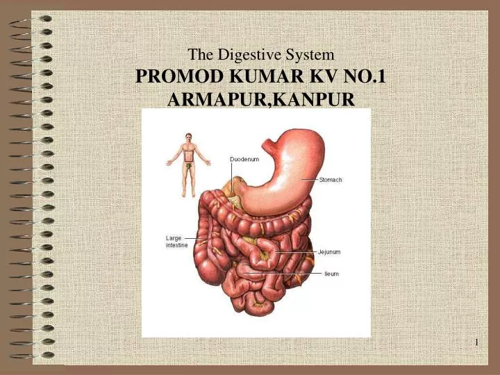
The Digestive System PROMOD KUMAR KV NO.1 ARMAPUR,KANPUR. Digestion. Processing of food Types Mechanical (physical) Chew Tear Grind Mash Mix Chemical Catabolic reactions Enzymatic hydrolysis Carbohydrate Protein Lipid. Digestion. Phases Ingestion Movement Digestion Absorption

E N D
Digestion • Processing of food • Types • Mechanical (physical) • Chew • Tear • Grind • Mash • Mix • Chemical • Catabolic reactions • Enzymatic hydrolysis • Carbohydrate • Protein • Lipid
Digestion • Phases • Ingestion • Movement • Digestion • Absorption • Further digestion
Digestive System Organization • Gastrointestinal (Gl) tract (Alimentary canal) • Tube within a tube • Direct link/path between organs • Structures • Mouth • Oral Cavity • Pharynx • Esophagus • Stomach • Duedenum • Jejenum • kIleum • Cecum • Ascending colon • Transverse colon
Descending colon Sigmoid colon Rectum Anus Accessory structures Not in tube path Organs Teeth Tongue Salivary glands Liver Gall bladder Pancreas Digestive System Organization
Sequence Voluntary stage Push food to back of mouth Pharyngeal stage Raise Soft palate Larynx + hyoid Tongue to soft palate Esophageal stage Contract pharyngeal muscles Open esophagus Start peristalsis Deglutition (swallowing)
Control Nerves Glossopharyngeal Vagus Accessory Brain stem Deglutition center Medulla oblongata Pons Disorders Dysphagia Aphagia Deglutition (swallowing)
Esophagus • Usually collapsed (closed) • 3 constrictions • Aortic arch • Left primary bronchus • Diaphragm • Surrounded by • SNS plexus • Blood vessels • Functions • Secrete mucous • Transport food
Esophagus • Sphincters • Upper • Lower • Abnormalities • Achalasia • Atresia • Hernia • Barret’s esophagus • Esophageal varices
Stomach • Usually “J” shaped • Left side, anterior to the spleen • Mucous membrane • G cells – make gastrin • Goblet cells – make mucous • Gastric pit – Oxyntic gland – Parietal cells – Make HCl • Chief cells – Zymogenic cells • Pepsin • Gastric lipase
3 muscle layers Oblique Circular Longitudinal Regions Cardiac sphincter Fundus Antrum (pylorus) Pyloric sphincter Vascular Inner surface thrown into folds – Rugae Contains enzymes that work best at pH 1-2 Stomach
Functions Mix food Reservoir Start digestion of Protein Nucleic acids Fats Activates some enzymes Destroy some bacteria Makes intrinsic factor – B 12 absorption Destroys some bacteria Absorbs Alcohol Water Lipophilic acid B 12 Stomach
Extends from pyloric sphincter ileocecal valve Regions Duodenum Jejenum Ileum Movements Segmentation Peristalsis Small Intestine
Small Intestine • Histology • Intestinal glands – Intestinal enzymes • Duodenal glands – Alkaline mucous • Paneth cells – Lysozyme • Microvilli • Lacteals • Plica circularis • Smooth muscle • Lymphatic tissue – GALT • Vascular
Absorbs 80% ingested water Electrolytes Vitamins Minerals Carbonates Active/facilitated transport Monosaccharides Proteins Di-/tripeptides Amino acids Lipids Monoglycerides Fatty acids Micelles Chylomicrons Small Intestine
Secretes digestive enzymes Peptidases Amino- Di- Tri- Sucrases Maltase Lactase Saccharidases Di- Tri- Lipase Nucleases Small Intestine
Control Requires pancreatic enzymes & bile to complete digestion Small Intestine
Large Intestine • Extends from ileocecal valve to anus • Regions • Cecum – Appendix • Colon • Ascending • Transverse • Descending • Rectum • Anal canal
Large Intestine • Histology • No villi • No permanent circular folds • Smooth muscle • Taeniae coli • Haustra • Epiploic appendages • Otherwise like rest of Gl tract
Large Intestine • Functions • Mechanical digestion • Haustral churning • Peristalsis • Reflexes • Gastroileal • Gastrocolic • Chemical digestion – Bacterial digestion • Ferment carbohydrates • Protein/amino acid breakdown • Absorbs • More water • Vitamins • B • K • Concentrate/eliminate wastes
Chyme dehydrated to form feces Feces composition Water Inorganic salts Epithelial cells Bacteria Byproducts of digestion Defecation Peristalsis pushes feces into rectum Rectal walls stretch Control Parasympathetic Voluntary Feces Formation and Defecation
Liver • Location • R. Hypochondrium • Epigastric region • 4 Lobes • Left • Quadrate • Caudate • Right • Each lobe has lobules – Contains hepatocytes – Surround sinusoids – Feed into central vein
Functions Makes bile Detergent – emulsifies fats Release promoted by: Vagus n. CCK Secretin Contains Water Bile salts Bile pigments Electrolytes Cholesterol Lecithin Liver
Detoxifies/removes Drugs Alcohol Stores Gycolgen Vitamins (A, D, E, K) Fe and other minerals Cholesterol Activates vitamin D Fetal RBC production Phagocytosis Metabolizes absorbed food molecules Carbohydrates Proteins Lipids Liver
Dual blood supply Hepatic portal vein Direct input from small intestine Hepatic artery/vein Direct links to heart Liver