Download
1 / 14
180 likes | 815 Views
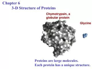
3-D Structure / Function. Myoglobin/ Hemoglobin. First protein structures determined Oxygen carriers Hemoglobin transport O 2 from lungs to tissues Myoglobin O 2 storage protein. Mb and Hb subunits structurally similar. 8 alpha-helices Contain heme group Mb monomeric protein

E N D
Myoglobin/Hemoglobin • First protein structures determined • Oxygen carriers • Hemoglobin transport O2 from lungs to tissues • Myoglobin O2 storage protein
Mb and Hb subunits structurally similar • 8 alpha-helices • Contain heme group • Mb monomeric protein • Hb heterotetramer (a2b2) myoglobin hemoglobin
Heme group • Heme = Fe++ bound to tertapyrrole ring (protoporphyrin IX complex) • Heme non-covalently bound to globin proteins through His residue • O2 binds non-covalently to heme Fe++, stabilized through H-bonding with another His residue • Heme group in hydrophobic crevice of globin protein
Oxygen Binding Curves • Mb has hyberbolic O2 binding curve • Mb binds O2 tightly. Releases at very low pO2 • Hb has sigmoidal O2 binding curve • Hb high affinity for O2 at high pO2 (lungs) • Hb low affinity for O2 at low pO2 (tissues)
O2 Binding to Hb shows positive cooperativity • Hb binds four O2 molecules • O2 affinity increases as each O2 molecule binds • Increased affinity due to conformation change • Deoxygenated form = T (tense) form = low affinity • Oxygenated form = R (relaxed) form = high affinity
T-conformation R-conformation O2 Binding induces conformation change Heme moves 0.34 nm Exposing crystal of deoxy-form to air cause crystal to crack
Allosteric Interactions • Allosteric interaction occur when specific molecules bind a protein and modulates activity • Allosteric modulators or allosteric effectors • Bind reversibly to site separate from functional binding or active site • Modulation of activity occurs through change in protein conformation • 2,3 bisphosphoglycerate (BPG), CO2 and protons are allosteric effectors of Hb binding of O2
Bohr Effect • Increased CO2 leads to decreased pH CO2+ H2O <-> HCO3-+ H+ • At decreased pH several key AA’s protonated, causes Hb to take on T-conformation (low affinty) • In R-form same AA’s deprotonated, form charge charge interactions with positive groups, stabilize R-conformation (High affinity) • HCO3- combines with N-terminal alpha-amino group to form carbamate group. --N3H+ + HCO3- --NHCOO- • Carbamation stabilizes T-conformation
Bisphosphoglycerate (BPG) • BPG involved acclimation to high altitude • Binding of BPG to Hb causes low O2 affinity • BPG binds in the cavity between beta-Hb subunits • Stabilizes T-conformation • Feta Hb (a2g2) has low affinity for BPG, allows fetus to compete for O2 with mother’s Hb (a2b2) in placenta.
Mutations in a- or b-globin genes can cause disease state • Sickle cell anemia – E6 to V6 • Causes V6 to bind to hydrophobic pocket in deoxy-Hb • Polymerizes to form long filaments • Cause sickling of cells • Sickle cell trait offers advantage against malaria • Fragile sickle cells can not support parasite