Download

1 / 44
570 likes | 960 Views
Preload Assessment. Too much of a good thing. Intravenous fluid boluses are used liberally in medicine to achieve a variety of goals. In critical care we use them most often to improve cardiovascular parameters and replace known losses

E N D
Too much of a good thing • Intravenous fluid boluses are used liberally in medicine to achieve a variety of goals. • In critical care we use them most often to improve cardiovascular parameters and replace known losses • Prompt, appropriate resuscitation with fluid can prevent the need for vasoactive drugs
Too much of a good thing • Excessive or inappropriate use of fluid can cause problems both acutely with APO or worsening of ALI and over the longer term with potentially delayed extubation, persistent oedema predisposing to infections or potential surgical complications like anastomotic breakdowns.
Preload assessment • Since it is harmful to give too much fluid as well as to give too little we need to have a good idea when its indicated. • When treating a cardiovascular parameter it is important to treat the patient not the numbers • E.g. Hypotension with no end organ dysfuncion • After establishing that the cardiac output/Blood pressure needs to be improved the next important question is: “Will this patient benefit from increased pre-load?”
What is pre-load? • Ventricular preload is the end-diastolic volume producing the initial passive stretch of the myocardium prior to active contraction. • Volume not pressure.
What is pre-load? • According to Starling ,One of 3 Determinants of Cardiac stroke volume • Pre-load • Sympathetic Tone • After-load
Why are we interested? • Increasing Pre-load increases Stroke volume. • Stroke volume x Heart rate = Cardiac Output • Cardiac output influences • Systemic Blood pressure • End organ perfusion • Oxygenation • Nutrients • Acid and waste removal • The answer to life the universe and everything
Frank and Starling • Cardiovascular Physiologists • Developed animal models where RA was canulated and supplied with blood from a resovior. Height of resovior determined pressure (Preload) • Aorta was attached to a variable resistor (Afterload) and fed back to resovior. • The Vagus and Sympathetic nerves could be artificially Stimulated. Image from P Cam “Physiology for the Anaesthetist”
Frank and Starling • The Ventricular volumes could then be measured. • These graphs show how increased end diastolic volume increased the stroke volume to a point then output was decreased as overstretch occurred. • Of note, these are volumes not pressures. The pressure curve has more of a plateau Image from P Cam “Physiology for the Anaesthetist”
Frank and Starling • This shows the same part of the curve we are interested in using, but substitues pressure as preload • Volume is proportional to pressure
Frank and Starling • Is it really that simple?
Guyton • The heart is a demand pump. • It can’t pump what it doesn’t receive from venous return. • In health the maximum Cardiac output achievable from solely increasing Preload is 10-15L/min • Adding sympathetic stimulation this increases to 20-30L/min • So there is usually some reserve Image from Current Opinion in Critical Care 2010, 16:289–296 “Fluid status and Fluid Responsiveness”
Guyton • Guyton’s venous return and cardiac output curves can be plotted simultaneously to show their point of intersection. • If you change the pre-load after a few cardiac cycles a new intersection point will be reached with a new cardiac output/venous return. • There is a plateau to the CO curve an increase in pre-load will not always work.
Preload assessment • The most important part about pre-load assessment is not what the pre-load currently is (CVP) but weather an increase will lead to a clinically significant effect. • The assessment of Pre-load and prediction of fluid responsiveness can be divided into Static and Dynamic measurements. • Each seeks to find a surrogate for the gold standard measurement of preload; the Left (or right) Ventricular End Diastolic volume (or pressure)
Static vs dynamic predictors • Static predictors seek to measure a single value in order to make a prediction on fluid responsiveness • Pressures CVP / Ppao • Volumes; Global end diastolic volume • Dynamic predictors apply a controlled and reversible preload variation and measure the heamodynamic response. • Variation with respiration: • Stroke volume Variation / Pulse Pressure variation • Plethysmographic variation • IVC variation • Systolic Pressure Variation • Passive leg raise test • Valsalva manoeuvre Ref: CritCare Clin 26 (2010) “Preload Assessment”
Static predictors • Pressures • CVP • Ppao • Volumes • Ventricular volumes (thermodilution) • Left Ventricular End Diastolic Area (U/S)
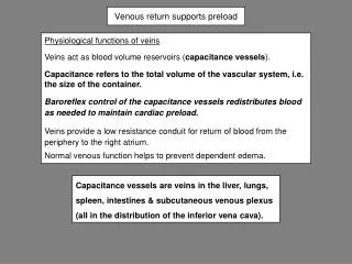
Static predictors • Central Venous Pressure • Gives you a surrogate for Left Ventricular end diastolic pressure. • Very unreliable • Needs to have a reproducible reference point to use absolute values (e.g. “CVP of less than 8 needs fluid”) • Stopcock of transducer needs to be zeroed level with right atrium. • Varies with Respiratory cycle, • Varies (Inconsistently) given different PEEP values • Totally unreliable if any right sided valvular disease.
Static predictors • Central Venous Pressure • Marikand Rodrigo did an update meta-analysis published in July this year By the Society of Critical Care Medicine • This reinforced that there was little to no correlation between CVP values and fluid responsiveness. Making a plea stating “this app to fluid resuscitation should be abandoned.” Ref: “Does the Central Venous Pressure Predict Fluid Responsiveness? An Updated Meta-Analysis and a Plea for Some Common Sense*” CritCareMed2013; 41:1774–1781
Static predictors • Pulmonary arterial occlusion pressure • Swan-Ganz catheter is passed into a branch of the pulmonary artery, a baloon inflated and the pressure beyond the balloon measured. • This pressure is thought to be proportional to Left atrial pressure and an idea of Left Ventricular preload gleaned much simmilar to using the static measurement of CVP. • It is an Invasive and potentially dangerous procedure and whilst a swan ganz is still the gold standard for measureing Cardiac output, performing Ppao has gone out of vouge Ref: “Static Measures of Preload Assessment” CritCare Clin 26 (2010) 295–305
Static predictors • Pulmonary arterial occlusion pressure • A 2002 review by Michard and Teboul of 12 studies found that Ppao could not reliably predict fluid responsiveness in any of the studies. • In general the best use for CVP or Ppao are in their negative predictive value. • A high CVP (e.g. >10 mm Hg in one study) is less likely to respond to a fluid bolus • A high Ppao (e.g. >11mm Hg in 2007 Osman Etal) is less likely to respond to a fluid bolus. Ref: “Static Measures of Preload Assessment” CritCare Clin 26 (2010) 295–305
Static predictors • Pressures • CVP • Ppao • Volumes • Ventricular volumes (thermodilution) • Left Ventricular End Diastolic Area (U/S)
Static predictors • Volumes • Right Ventricular End diastolic volume(thermodilution) • A Pulmonary artery catheter with a rapidly responsive thermister in the tip and a means of warming or cooling a known quantity and temperature of blood in the right atrium allows measurement of the SV, CO and RVEF. The RVEDV can then be derived from the SV and the RVEF. • Diebeletal. In 1994 compared RVEDVI in trauma patients to fluid responsiveness (20% increase in CI) and found that cut-offs of <90, 90-140, and >140 mL/m^2 had predictive values of 64%, 27% and 0% respectively.
Static predictors • Volumes • Global End Diastolic Volume (and index) • PiCCO, uses a Central line and a specially designed Femoral Arterial line to calculate, by thermo-dilution the GEDVI • Numerous studies exist showing good correlations between increases in GEDVI and SVI and one even compared these to CVPs which did not correlate to changes in SVI
Static predictors • In summary; • Pressures (CVP and Ppao) are only really useful to predict lack of response to fluids in the ICU and even that is subject to debate. • Volumes measured by a swan ganz catheter (RVEDV) or PiCCO (GEDVI) have a better correlation with fluid responsiveness than static Pressures but, by nature require specialised invasive monitoring.
Dynamic Predictors • Dynamic predictors apply a controlled and reversible preload variation and measure the heamodynamic response. • Variation with respiration: • Stroke volume Variation / Pulse Pressure variation • Plethysmographic variation • IVC variation • Systolic Pressure Variation • Passive leg raise test • Valsalva manoeuvre
Frank and Starling • For a constant Afterload and Sypathetic stimulation, An Increase in Pre-load will produce an increase in Stroke volume as described by this curve.
Frank and Starling • Small variations in pre-load, like those due to respiration can cause changes in the stroke volume or pulse pressure. • These small dynamic changes can be used to obtain an idea of the position on their Cardiac output curve and the potential for response to a fluid bolus.
Frank and Starling • During positive pressure ventilation inspiration right ventricular Preload is decreased due to IVC and RA compression leading to decreased venous return. • In turn this decreases RV output, Pulm Blood flow, LV Filling and LV output.
Frank and Starling • This also shows how static measures of pressure will be missleading in the setting of a failing ventricle. • The upper line represents a normal ventricle whilst the lower is failing, the same Pra representing dramatically different curve and potential for fluid responsiveness.
Dynamic Predictors • Variation with respiration: • Stroke volume Variation • Measured with a PiCCO or LiDCO via pulse contour analysis. • Shows promise as a predictor of fluid responsiveness but studies have given inconsistent results • Systolic Presure Variation • In it’s simplest for its a “swing” in the art line trace of a ventilated patient. • PreismanS et al showed that a variation of 8mmHg of systolic pressure correlates to fluid responsiveness with 82% sensitivity and 86% specificity • Preisman S et al Br J Anaesth2005;95:746–55
Dynamic Predictors • Variation with respiration: • Pulse Pressure Variation • Expressed as a percentage; PPV=100x(Ppmax-Ppmin)/[(Ppmax + PPmin/2] • More sensitive and specific than SPV. • Difference of 13% PPV would lead to at least 15% increase in Cardiac output with 500ml colloid (HES) (sens 94%, Spec 96%)
Dynamic Predictors • Variation with respiration: • Plethysmography assessed for “swing” similar to art line during respiration. Numerical value calculated usually giving a %change • Currently unreliable according to studies • Auto-gain in many systems dampens changes in the trace • Significant intra- and inter-patient variability. • No accepted standard or signal processing between manufacturers to facilitate reproduction between different institutions.
Dynamic Predictors • Variation with respiration: • IVC variation • The IVC pressure as it passes thru the diaphragm has nearly the same pressure as the Right Atrium. It also receives nearly all of the added pressure of Positive pressure ventilation. • During positive pressure inspiration the abdominal IVC expands as the abdominal pressure (usually) is significantly lower. This distension (and collapse) of the IVC only occurs in a fluid responsive patient. If it is full (i.e. Right heart failing) than ventilation will have little effect on IVC distension. • Using an Echo and M-mode the distension can be measured
Dynamic Predictors • Variation with respiration: • IVC variation • Barbier et al showed that if you calculate the dispensability as a percentage (Dmax-Dmin)/Dmin% if it was greater than 18% than it predicted fluid responsiveness with 90% sens and 90% spec.
Dynamic Predictors • Variation with respiration Caveats : • Must be Positive pressure ventilation, NOT triggering/PSV modes (I.e. Heavily sedated/narcotised, paralysed or just hyperventilated beyond pt’s resp drive) • Sinus Rhyth. • Limited by same things as Art Line (bubbles, kinks, excessive length etc) • Evidence of Fluid responsiveness does not equate to fluid Deficiency. • Limited data on extremes of ventilation E.g. Smaller tidal volumes with higher resp rates
Dynamic Predictors • Passive Leg Raising • Auto-transfusion from leg ~150-750 mLs depending on pt and technique. • Usable in Spontaneously ventilated patients. • Usable in Non-Sinus Rhythm. • Produces a change in CO within 30sec. Which resolves spontaneously (regardless of leg position due to blood re-distribution) • Most sensitive when pt starts semi-recumbent at 45deg then is laid flat and legs elevated. • Requires some means to rapidly assess changes in Cardiac output (usually TOE or TTE) • Needs more studies to make more easily clinically applicable.
Dynamic Predictors • Valsalvameneuver • Assessing primarily blood pressure trace as pt progresses thru the 4 phases of the Valsalvameneuver. • The presence of the variations in blood pressure, usually quantified using Pulse pressure Variation methods described earlier, with PPV of 52% or greater predicted fluid responsiveness. • Still inadequate studies, i.e. Difficult to justify using a Vigelo or PiCCO with such a simple bedside test.
Dynamic Predictors • Dynamic predictors apply a controlled and reversible preload variation and measure the heamodynamic response. • Variation with respiration: • Stroke volume Variation / Pulse Pressure variation • Plethysmographic variation • IVC variation • Systolic Pressure Variation • Passive leg raise test • Valsalva manoeuvre
In Summary • The Frank-Starling principle of ventricular stretch producing increased shortening/Stroke volume is key to assessing if a patient will respond to a fluid bolus • Healthy individuals will all respond to a fluid bolus! Just because a patient may respond doesn’t mean they need one. Use your clinical judgement on the adequacy of their cardiac output prior to assessing for responsiveness. • CVP only has some use as a predictor of NON-responsiveness • Dynamic measures ranging from Leg raise and Systolic pressure variation up to SVV and IVC distension are all potentially helpful tools in determining preload responsiveness. Each having their pros and cons. • Some potentially good research project ideas there!
Reference • Power and Kam, Principles of Physiology for the Anaesthetits 2e 2008 • “Dynamic Indicies of Preload” T. MikoEnomoto, Louise Harder, CritCare Clin 26 (2010) 307–321 • “Static Measures of Preload Assessment” Richard A. Nahouraii, Susan E. Rowell, Crit Care Clin 26 (2010) 295–305 • “Optimal volaemic status and predicting fluid responsiveness” Lorna Eyre, Andrew Breen Continuing Education in Anaesthesia, Critical Care & Pain | Volume 10 Number 2 2010 • “Fluid status and fluid responsiveness” Sheldon MagderCurrOpinCrit Care 16:289–296 • “Does the Central Venous Pressure Predict Fluid Responsiveness? An Updated Meta-Analysis and a Plea for Some Common Sense” Paul E. Marik, Rodrigo Cavallazzi, CritCare Med 2013; 41:1774–1781