Download
1 / 28
280 likes | 283 Views
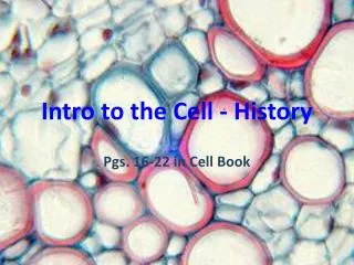
Intro to the Cell Ch. 6. Lecture Objectives 1. Cell History 2.Prokaryotic vs. Eukaryotic Cells 3. Cellular Organelles 4. Endosymbiont Theory. History of the Cell. 1. Robert Hooke a. first to name cells. 2. Van Leuwenhooke a. First to identify cells as living structures.

E N D
Intro to the Cell Ch. 6 Lecture Objectives1. Cell History 2.Prokaryotic vs. Eukaryotic Cells 3. Cellular Organelles 4. Endosymbiont Theory
History of the Cell 1. Robert Hooke a. first to name cells 2. Van Leuwenhooke a. First to identify cells as living structures
The Cell Theory: Schleiden & Schwann 1. All organisms are made of cells 2. The cell is the simplest collection of matter that can be alive 3. Cell structure is correlated to cellular function 4. All cells are related by their descent from earlier cells
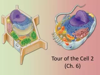
Comparing Prokaryotic & Eukaryotic Cells • Basic features of ALL cells a. Plasma membrane b. Semifluid substance = cytoplasm (area between PM and nucleus) c. Genetic Material d. Ribosomes e. cytoskeleton
Figure 6.8 Parent cell 1 μm 10 μm Cell wall Buds Vacuole Cell Animal Cells Fungal Cells Nucleus 5 μm Nucleolus Nucleus Yeast cells budding (colorized SEM) Human cells from lining of uterus (colorized TEM) Mitochondrion A single yeast cell (colorized TEM) Cell Flagella 1 μm 5 μm 8 μm Cell wall Chloroplast Nucleus Unicellular Eukaryotes Mitochondrion Nucleolus Plant Cells Nucleus Vacuole Nucleolus Chlamydomonas (colorized SEM) Chloroplast Cells from duckweed (colorized TEM) Cell wall Chlamydomonas (colorized TEM)
Prokaryotic cells are characterized by having ONLY 1. Cell Walls 2. Free-floating circular DNA 3. No membrane-bound organelles
Figure 6.5 Fimbriae Nucleoid Ribosomes Plasma membrane Bacterial chromosome Cell wall Glycocalyx 0.5μm Flagella (a) A typical rod-shapedbacterium (b) A thin section through thebacterium Corynebacteriumdiphtheriae (colorized TEM)
ENDOPLASMIC RETICULUM (ER) Figure 6.8 Nuclear envelope Rough ER Smooth ER NUCLEUS Nucleolus Flagellum Chromatin Centrosome Plasma membrane CYTOSKELETON: Microfilaments Intermediate filaments Microtubules Ribosomes Microvilli Golgi apparatus Peroxisome Lysosome Mitochondrion
Figure 6.8 Nuclear envelope NUCLEUS Nucleolus Rough ER Chromatin Smooth ER Ribosomes Central vacuole Golgi apparatus Microfilaments CYTOSKELETON Microtubules Mitochondrion Peroxisome Plasma membrane Chloroplast Cell wall Plasmodesmata Wall of adjacent cell
A. Nucleus i. Surrounded by two membranes = nuclear envelope, interrupted by pores ii. Houses genetic material (DNA)
Nucleus 1 μm Figure 6.9 Nucleus Nucleolus Chromatin Nuclear envelope: Inner membrane Outer membrane Nuclear pore Rough ER Pore complex Surface of nuclear envelope (TEM) Ribosome Close-up of nuclear envelope Chromatin 0.25 μm 0.5 μm Pore complexes (TEM) Nuclear lamina (TEM)
B. Endoplasmic Reticulum (ER) a. continuous with the nuclear envelope b. two distinct regions of ER • Smooth ER • synthesizes lipids • metabolizes carbohydrates • detoxifies drugs and poisons • Rough ER • manufactures proteins to be exported from cell
Figure 6.11 Smooth ER Rough ER Nuclear envelope Smooth ER Rough ER ER lumen Cisternae Transitional ER Ribosomes Transport vesicle 0.20 μm
C. Golgi apparatus a. flattened membranous sacs b. cis and trans sides c. Functions - Modifies products of the ER - Sorts & packages materials transport vesicle
Figure 6.12 Golgi apparatus cis face (“receiving” side of Golgi apparatus) 0.1 μm Cisternae trans face (“shipping” side of Golgi apparatus) TEM of Golgi apparatus
Fig. 6.15 Nucleus Rough ER Smooth ER cis Golgi Plasmamembrane trans Golgi
D. Lysosomesi. Membranous sac of digestive enzymes ii. Functions - autophagy/recycling - digestion
Figure 6.13 Vesicle containing two damaged organelles 1 μm Nucleus 1 μm Mitochondrion fragment Peroxisome fragment Lysosome Digestive enzymes Lysosome Lysosome Plasma membrane Peroxisome Digestion Food vacuole Digestion Mitochondrion Vesicle Phagocytosis: lysosome digesting food (b) (a) Autophagy: lysosome breaking down damaged organelles
E. Vacuoles i. diverse maintenance compartments (derived from ER & Golgi) ii. examples - food vacuoles - contractile vacuoles - central vacuoles
Figure 6.14 Central vacuole Cytosol Central vacuole Nucleus Cell wall Chloroplast 5 μm
Endomembrane System regulates protein traffic & performs metabolic functions • Components • Nuclear envelope • ER • Golgi apparatus • Lysosomes • Vacuoles • Plasma membrane • These components are either continuous or connected via transfer by vesicles
G. Mitochondria i. surrounded by a double membrane ii. possesses own DNA & ribosomes iii. Cellular respiration - # varies by cell type
Figure 6.17 Mitochondrion 10 μm Mitochondria Intermembrane space Outer membrane DNA Inner membrane Free ribosomes in the mitochondrial matrix Mitochondrial DNA Cristae Nuclear DNA Matrix 0.1 μm (a) Diagram and TEM of mitochondrion (b) Network of mitochondria in Euglena (LM)
H. Chloroplast i. stores chlorophyll - found in autotrophic organisms ii. photosynthesis - # varies by cell type
Figure 6.18 Chloroplast 50 μm Ribosomes Stroma Inner and outer membranes Granum Chloroplasts (red) DNA 1 μm Thylakoid Intermembrane space (a) Diagram and TEM of chloroplast Chloroplasts in an algal cell (b) (b)
The Endosymbiont theory 1. An early ancestor of eukaryotic cells engulfed a non-photosynthetic prokaryotic cell, which formed an endosymbiont relationship with its host 2. The host cell and endosymbiont merged into a single organism, a eukaryotic cell with a mitochondrion 3. At least one of these cells may have taken up a photosynthetic prokaryote, becoming the ancestor of cells that contain chloroplasts
Endoplasmicreticulum Nucleus Engulfing of oxygen-using nonphotosyntheticprokaryote, whichbecomes a mitochondrion Nuclear envelope Ancestor ofeukaryotic cells(host cell) Mitochondrion Engulfing ofphotosyntheticprokaryote At leastone cell Chloroplast Nonphotosyntheticeukaryote Mitochondrion Photosynthetic eukaryote