Download
1 / 52
520 likes | 557 Views
Clinical Approach to Thyroid Nodule. Mahtab Niroomand M.D. Assistant Professor of Endocrinology SBMU 1397/6/22. Agenda. Definition Epidemiology Clinical Importance Approach to thyroid nodule. Definition. Historically: palpable lump in the thyroid gland

E N D
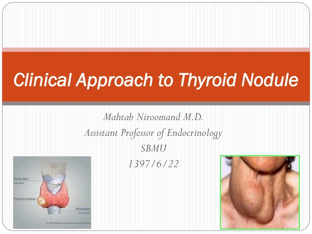
Clinical Approach to Thyroid Nodule MahtabNiroomand M.D. Assistant Professor of Endocrinology SBMU 1397/6/22
Agenda • Definition • Epidemiology • Clinical Importance • Approach to thyroid nodule
Definition • Historically: palpable lump in the thyroid gland • Now: discrete lesion within the thyroid gland that is radiologically distinct from the surrounding thyroid parenchyma. • Incidentaloma: • Nonpalpablenodules detected on US or other anatomic imaging studies are termed incidentally discovered nodules
Prevalence • Thyroid nodules are a common clinical problem. • Prevalence : • Studies based on inspection and palpation reported a 3 to 7% • Clinically inapparent thyroid nodules have been detected by US in 20% to as many as 76% of the general population a prevalence similar to that in autopsy data.
Prevalence • Palpable thyroid nodules in iodine-sufficient area : • 5% in women and 1% in men • Thyroid nodules are more common in: • Elderly persons, • Females, • Subjects from iodine-deficient geographic areas, • Those with a history of radiation exposure • Widespread use of imaging techniques has generated an epidemic of nonpalpable thyroid nodules
Clinical Importance • Both benign and malignant disorders can cause thyroid nodules • The clinical importance of a newly diagnosed thyroid nodule is primarily the exclusion of malignancy . • Thyroid cancer occurs in 7%–15% of thyroid nodules depending on: • Age • Sex • Radiation exposure history • Family history • ……..
Clinical Importance • DTC (differentiated thyroid cancers): • >90% of all thyroid cancers • Including : • Papillary • Follicular • Recent population-based study reported: • Doubling of thyroid cancer incidence from 2000 to 2012 compared to the prior decade
Management of thyroid Nodule • The basis of thyroid nodule management: • Clinical findings with: • Sensitive thyrotropin (TSH) assay • High-resolution ultrasonography (US) • Fine-needle aspiration (FNA) biopsy
Clinical findings : • History: • Age • Personal or family history of thyroid disease or cancer • Previous head or neck irradiation • Rate of neck mass growth • Anterior neck pain • Dysphonia, dysphagia, or dyspnea • Symptoms of hyper- or hypothyroidism • Use of iodine-containing drugs or supplements
Growth rate • Slow growth over years may be present in benignnodules • progressive growth during weeks or months may suggest malignancy. • The sudden appearance of a lump with pain: • Hemorrhage in a cystic nodule (more commonly) • Anaplastic thyroid carcinoma • Rare forms of chronic thyroiditis (Riedel disease) • Primary lymphoma of the thyroid
History • Symptoms such as : • Choking sensations • Vague cervical tenderness or pain • Dysphagia or • Hoarseness • The symptoms and signs of tracheal compression (cough and dysphonia) may suggest the risk of an underlying malignant lesion
Clinical findings : • Physical Examination: • Careful, focused examination of the thyroid gland and cervical lymph nodes: • Thyroid volume and consistency • Location, consistency, size, and number of nodule(s) • Neck tenderness or pain • Cervical adenopathy
Clinical findings : • Most nodules are asymptomatic and benign, but the absence of symptoms does not rule out malignancy [BEL 2, GRADE A]. • The risk of cancer is not substantially different in patients with a solitary nodule versus patients with a multinodular goiter (MNG) [BEL 2,GRADE B].
Sensitive TSH assay • Serum thyrotropin (TSH) should be measured during the initial evaluation of a patient with a thyroid nodule. (Strong recommendation, Moderate-quality evidence) • If the serum TSH is subnormal, a radionuclide (preferably 123I) thyroid scan should be performed. (Strong recommendation, Moderate-quality evidence) • If the serum TSH is normal or elevated, a radionuclide scan should not be performed as the initial imaging evaluation. (Strong recommendation, Moderate-quality evidence)
Sensitive TSH assay • Since hyperfunctioning nodules rarely harbor malignancy, no cytologic evaluation is necessary • Higher serum TSH level (even within the upper part of the reference range) is associated with increased risk of malignancy in a thyroid nodule, as well as more advanced stage thyroid cancer
Serum Thyroglobulin • Routine measurement of serum Tg for initial evaluation of thyroid nodules is not recommended. (Strong recommendation, Moderate-quality evidence)
Serum calcitonin • The panel cannot recommend either for or against routine measurement of serum calcitonin in patients with thyroid nodules. (No recommendation, Insufficient evidence) • May be considered in the subgroup of patients in whom an elevated calcitoninmay change the diagnostic or surgical approach: • Patients considered for less than total thyroidectomy • Patients with suspicious cytology not consistent with PTC • Unstimulatedserum calcitoninlevel >50–100 pg/mL: • Diagnosis of MTC is common
Thyroid Imaging • Thyroid sonography with survey of the cervical LN should be performed in all patients with known or suspected thyroid nodules. (Strong recommendation, High-quality evidence) • Ultrasound should evaluate the following: • Thyroid parenchyma (homogeneous or heterogeneous) • Thyroid gland size • Nodules size • Location • Sonographiccharacteristics of any nodule(s) • Ultrasound for FNA decision-making
Thyroid Ultrasonography: • When to Perform Thyroid Ultrasound: • US evaluation is recommended for: • Patients who are at risk for thyroid malignancy • Have palpable thyroid nodules or goiter • Have neck lymphadenopathy suggestive of a malignant lesion [BEL 2, GRADE A]. • US evaluation is not recommended : • As a screening test for the general population or patients with a normal thyroid on palpation and a low clinical risk of thyroid disease [BEL 4, GRADEC].
Thyroid Ultrasonography: • How to Describe US Findings: • Focus the US report on stratification for risk of malignancy • Describe position, size, shape, margins, content, echogenic pattern, and vascular features of the nodule(s) • For multiple nodules, detail the nodule(s) bearing the US characteristics associated with malignancy rather than describing the largest (dominant) nodule. • For suspicious regional neck lymph nodes, describe the cervical compartment, number, shape, size, margins, content, echogenic pattern, presence of hilum, and vascular features [BEL 2, GRADE A].
Comparison of the 2016 AACE/ACE-AME, 2015 ATA, and 2014 BTA Thyroid Nodule Ultrasound Classification Systems
Ultrasound Features of Lymph NodesPredictive of Malignant Involvement
Other Diagnostic Imaging Studies: • Radionuclide Scanning • When to Perform Thyroid Scintigraphy: • In a thyroid nodule or MNG, when the TSH level is below the lower limit of the reference range, or when ectopic thyroid tissue or a retrosternal goiter is suspected [BEL 2,GRADE A] • In iodine-deficient regions, to exclude autonomy of a thyroid nodule or MNG even when the TSH level is low-normal (e.g., 0.5-1.0 mIU/L). [BEL 3, GRADE B] AACE/ACE 2016
How to Perform Thyroid Scintigraphy • Use of 123I, 99mTcO4– (sodium pertechnetate), or technetium sestamibi can be considered for thyroid scintigraphy [BEL 3, GRADE C]. • Sodium iodide 131I thyroid uptake is not recommended for routine diagnostic use unless low-uptake thyrotoxicosis is suspected [BEL3, GRADE B].
Other Diagnostic Imaging Studies: • Thyroid lesions discovered on CT or MRI performed for other reasons have an uncertain risk of malignancy and should undergo US evaluation before considering FNA biopsy • Nodules detected by 18FDG PET have a high risk of malignancy when thyroid uptake is focal . • Such lesions should undergo a preliminary thyroid US evaluation because the presence of diffuse thyroid uptake may be linked to inflammatory conditions. • After confirmation of a true nodular lesion, FNA should be performed. • Focal lesions detected by 99mTc MIBI scans have a high risk of malignancy and should be evaluated carefully. • When MIBI is positive, the risk of malignancy in nodules is around 27%.
FNA • FNA is the procedure of choice in the evaluation of thyroid nodules, when clinically indicated. (Strong recommendation, High-quality evidence) • FNA: • Palpation-guide • US-guide
Palpation or US guide FNA • US-guided FNA is preferred for nodules with higher likelihood of : • Nondiagnosticcytology (>25%–50% cystic component) • Sampling error (difficult to palpate or posteriorlylocated nodules) • If the diagnostic US confirms the presence of a predominantly solid nodule corresponding to what is palpated, the FNA may be performed using palpation or US guidance.
Lymph Node Evaluation • Sonographicevaluation of the anterior cervical lymph node compartments (central and lateral) should be performed when thyroid nodules are detected . • FNA of the suspicious lymph node (for cytology and washout for Tg measurement) : • In US detects cervical lymph nodes that are sonographically suspicious for thyroid cancer
AACE/ACE Indication of FNA: • FNA is recommended for the following: • High US risk thyroid lesions ≥10 mm • Intermediate US risk thyroid lesions >20 mm • Low US risk thyroid lesions only when >20 mm and increasing in size or associated with a risk history and before thyroid surgery or minimally invasive ablation therapy [BEL 2,GRADE A] • FNA is not recommended for nodules that are: • Functional on scintigraphy [BEL 2, GRADE B].
FNA of thyroid incidentalomas • Manage thyroid incidentalomas according to the previously described criteria for nodule diagnosis [BEL 2, GRADE A]. • Perform US evaluation of incidentalomas detected by CT or MRI before consideration of FNA biopsy [BEL 2, GRADE A]. • We recommend that thyroid incidentalomas detected by PET with 18F-fluorodeoxyglucose (focal uptake, in particular) should undergo US evaluation and FNA because of the high risk of malignancy [BEL 2, GRADE A].
FNA of MNG and lymph nodes • We do not recommend the biopsy of more than 2 nodules in the same patient when the nodules are selected on the basis of the previously described criteria [BEL 3, GRADE C]. • If a radioisotope scan is available, we recommend not biopsying hot areas [BEL 2, GRADEB]. • In the presence of suspicious cervical lymphadenopathy, we recommend FNA for cytologic assessment of both the lymph node and the suspicious nodule(s) [BEL 2, GRADE A]. • We recommend the determination of thyroglobulin (Tg) or calcitonin, according to clinical indications, on FNA washout of suspicious lymph nodes [BEL 2, GRADE A].
FNA of complex thyroid nodule(s) • We recommend sampling the solid component of the lesion through FNA biopsy[BEL 3,GRADE B]. • Preferentially sample the vascularized areas of complex lesions [BEL 4, GRADE C]. • Submit both the FNA specimen and the drained fluid for cytologic examination [BEL2, GRADE A].
FNA interpretation • Thyroid nodule FNA cytology should be reported using diagnostic groups outlined in the Bethesda System for Reporting Thyroid Cytopathology. (Strong recommendation, Moderate-quality evidence)
FNA reports: • Criteria for cytologic adequacy : • Presence of at least six groups of well-visualized follicular cells, each group containing at least 10 well-preserved epithelial cells, preferably on a single slide)
Long term follow up of thyroid Nodule • Follow-up of thyroid nodules with benign cytology diagnoses should be determined by risk stratification based upon US pattern. • Nodules with high suspicion US pattern: repeat US and US-guided FNA within 12 months. (Strong recommendation, Moderate-quality evidence)
Long term follow up of thyroid Nodule • Nodules with low to intermediate suspicion US pattern repeat US at 12–24 months. • If sonographic evidence of growth (20% increase in at least two nodule dimensions with a minimal increase of 2mm or more than a 50% change in volume) or development of new suspicious sonographic features, the FNA could be repeated or observation continued with repeat US, with repeat FNA in case of continued growth. (Weak recommendation, Low-quality evidence)
Long term follow up of thyroid Nodule • Nodules with very low suspicion US pattern (including spongiform nodules): the utility of surveillance US and assessment of nodule growth as an indicator for repeat FNA to detect a missed malignancy is limited. • If US is repeated, it should be done at >24 months. (Weak recommendation, Low-quality evidence)
Follow-up of nodules with two benign FNA cytology results • If a nodule has undergone repeat US-guided FNA with a second benign cytology result: • US surveillance for this nodule for continued risk of malignancy is no longer indicated. (Strong recommendation, Moderate-quality evidence)
Follow-up for nodules that do not meet FNA criteria • The strategy for sonographic follow-up of nodules that do not meet criteria for FNA at initial imaging should be based upon the nodule’s sonographic pattern: • Nodules with high suspicion US pattern: repeat US in 6–12 months. (Weak recommendation, Low-quality evidence) • Nodules with low to intermediate suspicion US pattern: consider repeat US at 12–24 months. (Weak recommendation, Low-quality evidence)