Download
1 / 31
320 likes | 1.56k Views
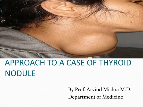
Case present. A 42-year-old woman presents with a palpable mass on the left side of her neck.She has no neck pain and no symptoms of thyroid dysfunction.Physical examination reveals a solitary, mobile thyroid nodule, 2 cm by 3 cm, without lymphadenopathy.. . The patient has no family history of th

E N D
1. The Thyroid Nodule ???
96/05/04
N Eng J Med 2004;351:1764-71.
Laszlo Hegedus, M.D.
2. Case present A 42-year-old woman presents with a palpable mass on the left side of her neck.
She has no neck pain and no symptoms of thyroid dysfunction.
Physical examination reveals a solitary, mobile thyroid nodule, 2 cm by 3 cm, without lymphadenopathy.
3. The patient has no family history of thyroid disease and no history of external irradiation.
Which investigations should be performed?
Assuming that the nodule is benign,which, if any, treatment should be recommended?
4. The clinical problem In the United States, 4 to 7 percent of the adult population have a palpable thyroid nodule.
Nodules are more common in women and increase in frequency with age and with decreasing iodine intake.
The prevalence is much greater with the inclusion of nodules that are detected by ultrasonography or at autopsy.
Malignant nodule corresponds to approximately 2 to 4 per 100,000 people per year, constituting only 1 percent of all cancers and 0.5 percent of all cancer deaths.
5. The clinical spectrum ranges from the incidental, asymptomatic, small, solitary nodule, in which the exclusion of cancer is the major concern, to the large, partly intrathoracic nodule that causes pressure symptoms, for which treatment is warranted regardless of cause.
The most common diagnoses and their approximate distributions are colloid nodules, cysts, and thyroiditis (in 80 percent of cases); benign follicular neoplasms (in 10 to 15 percent); and thyroid carcinoma (in 5 percent).
6. This review will focus on the management of a solitary thyroid nodule that is detected on physical examination, regardless of the finding of additional nodules by radionuclide scanning or ultrasonography, since such a finding does not alter the risk of cancer.
7. History and physical examination The history and physical examination remain the diagnostic cornerstones in evaluating the patient with a thyroid nodule and may be suggestive of thyroid carcinoma.
However, a minority of patients with malignant nodules have suggestive findings, which often also occur in patients with benign thyroid disorders.
9. The risk of thyroid cancer seems nearly as high in incidental nodules (<10 mm), the majority of which escape detection by palpation, as in larger nodules.
However, the vast majority of these microcarcinomas do not grow during long-term follow-up and do not cause clinically significant thyroid cancer.
The fact that ultrasonography detects nodules (a third of which are more than 20 mm in diameter) in up to 50 percent of patients with a normal neck examination underscores the low specificity and sensitivity of clinical examination.
10. When two or more risk factors that indicate a high clinical suspicion are present, the likelihood of cancer approaches 100 percent.
In such cases, biopsy is still useful to guide the type of surgery.
12. Laboratory investigations Because clinical examination is not sensitive for identifying thyroid dysfunction, laboratory evaluation of thyroid function is routinely warranted.
The only biochemical test routinely needed is measurement of the serum thyrotropin level.
If this level is subnormal, levels of free thyroxine or free triiodothyronine should be measured to document the presence and degree of hyperthyroidism.
13. Approximately 10 percent of patients with a solitary nodule have a suppressed level of serum thyrotropin, which suggests a benign hyperfunctioning nodule.
If the serum thyrotropin concentration is elevated, a serum antithyroperoxidase antibody level should be obtained to confirm Hashimoto�s thyroiditis.
If a patient has a family history of medullary thyroid cancer or multiple endocrine neoplasia type 2, a basal serum calcitonin level should be obtained; an elevated level suggests medullary thyroid cancer.
Before surgery is performed, investigation for primary hyperparathyroidism and pheochromocytoma should be carried out.
14. Imaging of the thyroid nodule Radionuclide scanning, which is performed much more commonly in Europe than in the United States, may be used to identify whether a nodule is functioning.
A functioning nodule, with or without suppression of extranodular uptake, is nearly always benign, whereas a nonfunctioning nodule, constituting approximately 90 percent of nodules, has a 5 percent risk of being malignant.
16. Thus, in the patient with a suppressed level of serum thyrotropin, radionuclide confirmation of a functioning nodule may obviate the need for biopsy.
A scan can also indicate whether a clinically solitary nodule is a dominant nodule in an otherwise multinodular gland and can reveal substernal extension of the thyroid.
17. Ultrasonography can accurately detect nonpalpable nodules, estimate the size of the nodule and the volume of the goiter, and differentiate simple cysts, which have a low risk of being malignant, from solid nodules or from mixed cystic and solid nodules, which have a 5 percent risk of being malignant.
Ultrasonography also provides guidance for diagnostic procedures (e.g., fine-needle aspiration biopsy) as well as therapeutic procedures (e.g., cyst aspiration, ethanol injection, or laser therapy) and facilitates the monitoring of the effects of treatment.
19. Characteristics revealed by ultrasonography � such as hypoechogenicity, microcalcifications, irregular margins, increased nodular flow visualized by Doppler, and, especially, the evidence of invasion or regional lymphadenopathy � are associated with an increased risk of cancer; however, sonographic findings cannot reliably distinguish between benign and cancerous lesions.
20. Fine-needle aspiration biopsy Independent of morphology, fine-needle aspiration provides the most direct and specific information about a thyroid nodule.
Complications are rare and primarily involve local discomfort.
Fine-needle aspiration has diagnostically useful results in about 80 percent of cases, typically with two to four passes of the needle.
The diagnostic accuracy of fine-needle aspiration depends on the way in which suspicious lesions are handled.
21. If fine-needle aspiration reveals a follicular neoplasm (which occurs in approximately 15 percent of nodules, of which only 20 percent turn out to be malignant), radionuclide scanning should be performed.
If such scanning shows a functioning nodule with or without complete suppression of the rest of the thyroid, surgery can be avoided, since the risk of cancer is negligible.
In a cystic lesion or one that is a mixture of cystic and solid components, fine-needle aspiration of a possible solid component should be performed, since the risk of cancer is the same as that for a solid nonfunctioning nodule.
22. Treatment of the solitary thyroid nodule The natural history of solitary thyroid nodules is poorly understood, mainly because nodules that are suspicious for cancer, cause pressure, or prompt reports of cosmetic problems are rarely left untreated.
With this reservation, it seems that the majority of benign nonfunctioning nodules grow, particularly those that are solid.
In one study, 89 percent of nodules that were followed for five years increased by 15 percent or more in volume.
23. The annual rate of evolution of a solitary functioning nodule into a hyperfunctioning nodule is as high as 6 percent; the risk is positively related to the size of the nodule and negatively related to the serum thyrotropin level.
There is controversy as to whether a solitary nodule should be treated and, if so, how.
Table 2 summarizes the advantages and disadvantages of potential treatment options.
25. Guidelines from professional societies Clinical-practice guidelines were published in 1996 by the American Thyroid Association(www.thyroid.org/professionals/publications/guidelines.html) and the American Association of Clinical Endocrinologists (www.aace.com/clin/guidelines/thyroid_nodules.pdf ).
26. Radionuclide scanning is not routinely recommended, but it is advocated in the case of a suppressed level of serum thyrotropin or the finding of follicular neoplasia with the use of fine-needle aspiration.
Thyroid ultrasonography is recommended to guide fine-needle aspiration, especially in nodules that are small and incidental or are partly cystic or from which primary fine-needle aspiration has yielded insufficient material.
Fine-needle biopsy of all possibly malignant nodules (which are not defined in the guidelines) is advocated.
27. In the case of a benign nodule, periodic lifelong follow-up every 6 to 24 months (including measurement of serum thyrotropin levels, neck palpation, and fine-needle aspiration in case of growth or other suspicious signs) is recommended.
For a functioning benign nodule, iodine-131 is considered the treatment of choice, with surgery as an alternative, especially if the nodule is very large or partly cystic or if the patient is young; treatment is more strongly recommended if the serum thyrotropin level is decreased or overt hyperthyroidism is present, because of adverse effects on bone and the cardiovascular system.
28. For a nonfunctional benign nodule, there is no clear recommendation on the use of levothyroxine, although this therapy is considered contraindicated when the serum thyrotropin level is suppressed, in patients more than 60 years old, and in postmenopausal women.
If levothyroxine therapy is used, regular reassessment (the interval is not defined in the guidelines) is recommended, with monitoring of serum thyrotropin levels, which should be subnormal but measurable.
29. Conclusions and recommendations For the patient who presents with a nodule, as in the case described in the vignette, the main concern is to exclude the possibility of thyroid cancer,even though the vast majority of nodules are benign.
The initial evaluation should include measurement of the serum thyrotropin level and a fine-needle aspiration, preferably guided by ultrasonography.
If the patient has a family history of medullary thyroid carcinoma or multiple endocrine neoplasia type 2, the serum calcitonin level should also be checked.
30. If the thyrotropin level is suppressed, radionuclide scanning should be performed.
In patients less than 20 years old, and in the case of a high clinical suspicion for cancer (e.g.,follicular neoplasia as diagnosed by fine-needle aspiration and a nonfunctioning nodule revealed on scanning), the patient should be offered hemithyroidectomy regardless of the results of fine-needle aspiration.
In the case of a functioning benign nodule, iodine-131 is generally the therapy of choice, independent of concomitant hyperthyroidism.
31. For nonfunctioning cystic nodules, aspiration and ethanol injection therapy may be considered, and ethanol injection or laser therapy if the nodules are solid, but data to support the use of these therapies are limited.
My usual approach after documenting benign cytology is to follow the patient yearly with neck palpation and measurement of the serum thyrotropin level, with repeated ultrasonography and fine-needle aspiration if there is evidence of growth of the nodule.
I do not recommend levothyroxine therapy to shrink or prevent growth of benign nodules because of the drug�s low efficacy and potential side effects.
32. The End Thank you for your attention.