Download
1 / 22
220 likes | 298 Views
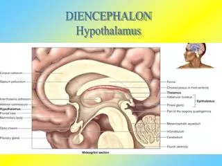
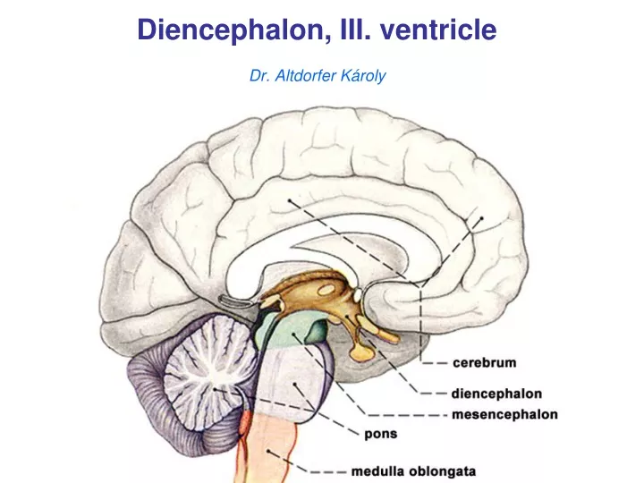
Diencephalon , III. ventricle Dr. Altdorfer Károly. Diencephalon. Thalamus Epithalamus Metathalamus Subthalamus Hypothalamus. Diencephalon. Thalamus Epithalamus habenulae habenular nuclei striae medullaris thalami habenular trigone habenular commissurre pineal body Metathalamus

E N D
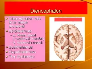
Diencephalon • Thalamus • Epithalamus • Metathalamus • Subthalamus • Hypothalamus
Diencephalon • Thalamus • Epithalamus • habenulae • habenular nuclei • striae medullaris thalami • habenular trigone • habenular commissurre • pineal body • Metathalamus • medial geniculate body • lateral geniculate body • Subthalamus • subthalamic nucleus (Luys) • zona incerta • Forel- H fields • Hypothalamus
I- II. III. IV.
THIRD VENTRICLE Reaching it from above
THIRD VENTRICLE: small, narrow, midline vertical cleft of diencephalon, bridged by interthalamic adhesion of thalamus. Roof:choroid lamina epithelialis of the 3rd venticle (+ choroid tela and choroid plexus). The attachments of choroid lamina epithelialis: tenia thalami (on stria medullaris thalami), habenulae and habenular commissure.
THIRD VENTRICLE: lateral wall
THIRD VENTRICLE: • Lateral wall: thalamus, hypothalamic sulcus and hypothalamus. • Floor: hypothalamus (optic and infundibular recesses). • Anterior wall: anterior commissure, columns of fornix and lamina terminalis (triangular recess). • Posteriorly: habenular commissure, posterior commissure (between them the pineal recess, above the habenular comm. the suprapineal recess). • OPENINGS: 2 interventricular foramina laterally, cerebral aqueduct posteriorly.
Posteriorly: habenular commissure, posterior commissure (between them the pineal recess, above the habenular comm. the suprapineal recess).
THIRD VENTRICLE: small, narrow, midline vertical cleft of diencephalon, bridged by interthalamic adhesion of thalamus. Lateral wall: thalamus, hypothalamic sulcus and hypothalamus. Roof:choroid lamina epithelialis of the 3rd venticle (+ choroid tela and choroid plexus). The attachments of choroid lamina epithelialis: tenia thalami (on stria medullaris thalami), habenulae and habenular commissure. Floor: hypothalamus (optic and infundibular recesses). Anterior wall: anterior commissure, columns of fornix and lamina terminalis (triangular recess). Posteriorly: habenular commissure, posterior commissure (between them the pineal recess, above the habenular comm. the suprapineal recess). OPENINGS: 2 interventricular foramina laterally, cerebral aqueduct posteriorly.
Lateral ventricles and III. ventricle
Central part of the lateral ventricle and the third ventricle (coronal section) 1. Corpus callosum 2. Fornix (body) 3. Tela choroidea with choroid plexus 4. Choroid lamina epithelialis 5. Central part of lat. ventr. 6. Stria terminalis+thalamostriate vein 7. Thalamus+hypothalamus 8. Third ventricle 9. Choroid lamina epithelialis of third ventr. 10. Tela choroidea with choroid plexus 11. Lamina affixa thalami 12. Body of caudate nucl.
Circumventricular organs organ
Cerebral fitness Use it – or lose it!