Download
1 / 1
10 likes | 113 Views
Delayed left ventricular relaxation due to smoking in healthy subjects: a strain-imaging study K. Farsalinos , D. Tsiapras , S . Kyrzopoulos , E . Avramidou , D . Vassilopoulou , V . Voudris

E N D
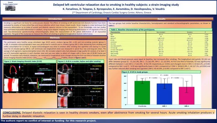
Delayed left ventricular relaxation due to smoking in healthy subjects: a strain-imaging study K. Farsalinos, D.Tsiapras, S. Kyrzopoulos, E. Avramidou, D. Vassilopoulou, V. Voudris 2nd Department of Cardiology, Onassis Cardiac Surgery Center, Athens, Greece Onassis Cardiac Surgery Center Introduction Results The two groups had similar baseline characteristics, hemodynamic and standard echocardiographic parameters, as shown in table 1. Heart rate and blood pressure were equal at baseline, but increased after smoking. The longitudinal end-systolic GS did not differ between groups (C: -21.2±1.9%, SM-1: -21.2±2.6%, SM-2: -21.1±2.8%). At first one-third of diastole, GS was significantly different between C and SM-1 (-7.2±1.6%, -8.7±2.5% respectively, p= 0.001) and changed more in SM-2 (-10.6±3.2%, p<0.001 compared to SM-1 and C). SI-DI was significantly lower in SM-1 compared to C (SM-1: 58.8±13.8%, C: 66.1±7.3%, p=0.004) and was further reduced after smoking two cigarettes (SM-2: 49.4±16%, p<0.001 compared to SM-1 and C). Smoking is a significant risk factor for cardiovascular disease. The effects of smoking on left ventricular (LV) diastolic function have been evaluated by studies of trans-mitral flow and tissue velocities which have shown mixed results.Strain imaging is a new technique that measures deformation as an index of systolic and diastolic capacity intrinsic to the myocardium. To our knowledge, only two studies have estimated myocardial deformation in smokers, using tissue doppler imaging and measuring deformation in 3-4 segments of the LV wall. Two-dimensional speckle-tracking echocardiography allows the measurement of the global deformation of all myocardial segments. The goal of this study was to measure the effects of smoking on global LV longitudinal diastolic deformation. Diastolic time 1/3 of diastole Table 1.Baseline characteristics of the participants. GSend-systole Methods The participants were healthy young volunteers (age 18-45 years), smokers (group SM, n=42) and non-smoking controls (group C, n=38). A complete echocardiographic exam was performed in controls and in smokers (group SM-1) after abstaining from smoking and coffee consumption for 12 hours. A repeat echocardiogram was done in smokers after smoking two cigarettes and staying in a quiet room for 15 minutes (group SM-2). Left ventricular (LV) longitudinal strain was measured in apical four, two and long-axis views. The average of all segments represented global strain (GS). We included subjects who had adequate image quality for analysis of at least 5 out of 6 myocardial segments in each chamber viewand care was taken to obtain all images with a frame rate of 60-80 frames per second. To examine early diastolic relaxation, we measured LV longitudinal strain changes during the first one-third of diastole duration (Strain Imaging-Diastolic Index [SI-DI] = [(GSend-systole – GS1/3-diastole)/GSend-systole]*100%), shown in figure 1 and 2. Figure 1.Strain Imaging-Diastolic Index (SI-DI). Figure 2.SI-DI in a smoker, before and after smoking Figure 3.SI-DI in study groups. CONCLUSIONS.Delayed diastolic relaxation is seen in healthy chronic smokers, even after abstinence from smoking for several hours. Acute smoking inhalation produces a further delay in diastolic relaxation. The authors report no conflict of interest or funding for this research project.