Download
1 / 29
290 likes | 433 Views
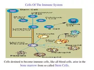
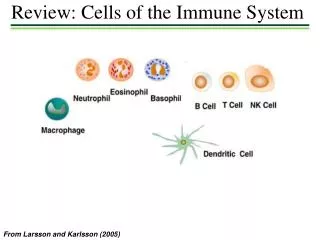
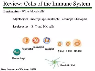
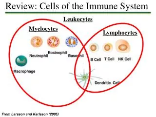
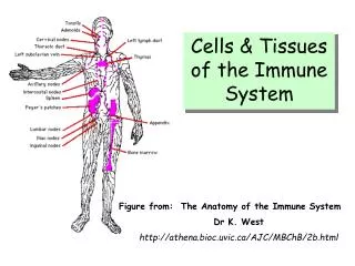
بسم اللة الرحمن الرحيم. Cells of Immune System. Cells of Immune System. Stem cells of bon marrow differentiate into cytokines (IL-&, IL-3) colony stimulating factor Lymphoid series Myeloid series B-lymphocytes T-lymphocytes NK

E N D
Cells of Immune System Stem cells of bon marrow differentiate into cytokines (IL-&, IL-3) colony stimulating factor Lymphoid series Myeloid series B-lymphocytes T-lymphocytes NK monocytee-macrophages dendritic cells eosinophils mast cells
The Life Of The B Cell • B lymphocytes are formed within the bone marrow and undergo their development there They have the following functions: • To interact with antigenic epitopes, using their immunoglobulin receptors • To subsequently develop into plasma cells, secreting large amounts of specific antibody, or • To circulate as memory cells • To present antigenic peptides to T cells, consequent upon interiorization and processing of the original antigen
* B cells become plasma cells, which produce antibodies when a foreign antigen triggers the immune response
B-lymphocytes in bon marrow * The lymphoid stem cells differentiate into B cells * B-cells precursors mature, differentiate into immunocomptent B-cells with a single antigen specificity * Immature B-cells that express high affinity receptors for self antigens, die or fail to mature i.e negative selection or clonal deletion * This process induces central self tolerance and reduces autoimmune diseases
B-lympocytes * Immature B cells express IgM receptors on the surface * Mature B cells express IgM, IgD molecules on surfaces * IgM and IgD molecules serve as receptors for antigens * Memory B-cells express IgG or IgA or IgE on the surface * B-cells bear receptors for Fc portion of IgG and a receptor for C3 component of the complement * They express an array of molecules on their surfaces that are important in B-cells interactions with other cells such as MHC II, B7 and CD40
Mechanism of Humoral immunity * Antibodies induce resistance through: 1) Antitoxin neutralize bacterial toxins (diphtheria,tetanus) Antitoxin are developed actively as a result of: a- Previous infection b- Artificial immunization c- Transferred passively as antiserum * Neutralization of toxin with antitoxin prevents a combination with tissue cells
Mechanism of Humoral immunity 2) Antibodies attach to the surface of bacteria and a- act as opsonins and enhance phagocytosisd b- prevent the adherence of microorganisms to their target cells, e.g. IgA in the gut c- Activate the complement and lead to bacterial lysis d- Clump bacteria (agglutination) leading to phagocytosis
T-Lmphocytes T-lmphocytes migrate from bon marrow to enter thymus 1) In the outer cortex of thymus: - T-lymphocytes acquire specific receptors (TCRs) - This receptor commit lymphocyte to a single antigen specificty - Responding by proliferation and production of a clone of cells (clonal selection) - They differentiate to express CD3, both CD4 and CD8 coreceptors (double positive cells)
* T lymphocytes become CD4+ (helper T cells) or* CD8+ cells (which in turn can become killer T cells) also called cytotoxic T cells
T-Lmphocytes 2) In the medulla of thymus: - TCRs recognize MHC molecules, loaded with normal self-peptides (p-MHC) - TCRs capable of binding with low affinity to p-MHC will receive positive selection signals to divide and establish clones - TCRs that bind too strongly to p-MHC undergo (negative selection) - This selection process will eliminate the potentially most harmful self reactive T-cells (central self tolerance)
T-Lmphocytes 3)Immature T-cells express both CD4 and CD8 (DP) As they mature * T-cell with TCRs that have affinity to bind to MHC class II will become helper T-cells with CD4 molecule only * T-cell with TCRs that have affinity to bind with MHC class I will become cytotoxic T-cells with CD8 molecule only
T-Lmphocytes 4) Mature positively selected T-cells are MHC restricted * CD4 T-cells are MHC II restricted and only recognize specific foreign peptide only when they are presented in association with specific MHC II molecules * CD8 T-cells are MHC I restricted and recognize specific foreign peptidees only when they are presented in association with specific MHC I molecules
T-cell surface markers These are molecules that by witch we can identify T-cells and divide them to subsets They are required to for interactions between T-cells and APC and for antigen recognition These are TCRs, CD3, CD4, CD8, CD2, CD28,and CD40 on activated T-cells
T-cell subpopulation 1) CD4 T helper lymphocytes (TH) - TH lymphocytes recognize antigen on the surface of APC in association with class II MHC molecules - They are activated and secrete several cytokines - There are two main subsets of TH cells (THI and TH2) - The two subsets are differentiated on basis of the cytokine they produce
1) CD4 T helper lymphocytes Subsets Th1 produce mainly : - Cytokines of CMI and inflammation e.g. IFN-γ, TNF- β, IL-3 and IL-2 TH2 produce mainly: - Cytokines that stimulate B-cells - Suppressor cytokines e.g. Il-4, IL-5, IL-6 and IL-10
2) CD8 Cytotoxic T-lymphocytes (CTLs) * They constitute 35% 0o peripheral T-cells * CTLs recognize antigen on suurface of target cells (infected APC or other infected nucleotid cell) in association with MHC-I * They are activated and kill the virus infected cell or tumour cell
Professional APCs Dendritic cells, macrophages, and B-lymphocytes Dendritic cells: - They are the most efficient APCs - They are the main inducers of primary immune response - Presenting antigen to and activating native T-cells in the recognition phase - They express class I and class II MHC molecules - Dendritic cells are primarly located under skin and mucosa of most organs - They capture foreign antigens and transport them to local lymph nods - They present antigen to native helper T-cells
Macrophages * Derived from myeloid stem cells in bon marrow * They exist as free cells in blood e.g. monocytes and fixed cells in tissues e.g. Kupffer cells of liver * They are important link between innate and aquired immune responses * They are activated and attracted to the site of foreign material by action of different cytokines e.g IFN-γ , C5a
Functions of Macrophages 1) Pagocytosis 2) Opsonization 3) APCs: they ingest foreign material, process it, and fragments of antigen are presented on its surface (in association with MHC molecules) for interaction with T-cells 4) Macrophages may kill antibody coated infected cells or tumour cells through release of lytic enzymes 5) They produce IL-1, IL-6, IL-12, IL-15, TNF-alpha 6) They secret prostaglandins and synthesize complement compononts
Natural killer (NK) Cells * Large granular lymphocytes which lack most surface markers of B and T-cells * They comprise 5-10% of the peripheral lymphocytes * They function mainly in innate immunity * They have spontaneous non-specific cytotoxic activity on virus infected cells, tumour cells and graft cells * They are not MHC restricted and MHC I inhibits their killing functions * The mechanism of NK mediated cytolysis is as that of CTLs
NK cells differ from CTLs in 1)They are non-specific 2)They act spontaneously without prior recognition or activation 3)They do not require antigen presentation by MHC 4)They destroy cells coated with antibodies, a mechanism called antibody dependant cellular cytotoxicity (ADDCC)
Antibodies produced by B-cells of the immune system recognize foreign antigens and mark them for destruction
Primary And Secondary Response • Primary Response: • Slow in Onset • Low in Magnitude • Short Lived • IgM • Secondary Response: • Rapid in Onset • High in Magnitude • Long Lived • IgG (Or IgA, or IgE