Download
1 / 105
1.28k likes | 1.82k Views
Tumors of Larynx. Prepared by Dr.Hiwa Asad. Benign Tumors. Ectodermal Mesodermal pseudotumours. Ectodermal Tumours. Papilloma: single or multiple Adenoma Paraganglioma Neurilemmoma. Single papilloma. Common in adults, rare in children Sessile or pedunculated

E N D
Tumors of Larynx Prepared by Dr.Hiwa Asad
Benign Tumors • Ectodermal • Mesodermal • pseudotumours
Ectodermal Tumours • Papilloma: single or multiple • Adenoma • Paraganglioma • Neurilemmoma
Single papilloma • Common in adults, rare in children • Sessile or pedunculated • Usual sites anterior commissure, anterior half of the vocal cords • Men : women ratio 2:1 • Present with hoarseness • If small removed endoscopically • If large by laryngofissure • Biopsy to exclude malignancy specially if recurrent
Squamous papilloma of the aryepiglottic fold Laryngeal papilloma
Multiple papillomas • Infants and young children, rare in adults • A virus may be responsible (HPV) • Vocal cords are the usual site • Hoarsness if vocal cords affected • Dyspnoea may occur ---- tracheostomy • Removed endoscopically by CO2 laser • Spontaneous recovery in puberty may occur
Juvenile papillomas Before and after removal
Mesodermal tumours • Vascular neoplasms • Chondroma • Myogenic tumours • Fibroma • Lipoma
Vascular neoplasms • Arise from blood or lymphatic vessels Haemangioma • Rare in adults • Telangiectatic vocal cord polyp
Chondroma • Arise from cartilages (Mostly cricoid) • More in men (40-70 years) Clinical features • Hoarsness and dyspnoea • Stridor (extention into subglottic space) • Dysphagia (extension into hypopharynx) • External swelling (cricoid ring or thyroid cartilage)
Chondroma • Indirect laryngoscopy reveals a smooth mass covered by intact mucosa • Radiology shows calcific stippling or coarse irregular calcificatuon • Biopsy specimens is unrepresentative, the tumour is hard and difficult to penetrate • Surgery is the treatment of choice • Radiptherapy is of little value
Malignant Tumors • 1% of all malignancies In UK • More in men • Predominantly of squamous pathology • Interfere with function and emotion • High cure rate 85%
Incidence • Higher in urban than rural population • Social and racial differences reflect different habits (tobacco and alcohol)
Classification The International Union against Cancer (UICC) classified Ca larynx on anatomical bases 20% 10% 70%
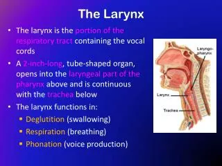
Epilarynx Suprahyoid epiglottis Aryepiglottic folds Supraglottis infrahyoid epiglottis false cords ventricles Supraglottis Glottis True cord,ant&postcomissure 1 cm Subglottis UICC classification of Ca larynx
Glottis true cords anterior commissure posterior commissure
Aetiology • Unknown • Possibly related factors genetic and social factors male predominance racial predilection urban pollution tobacco and alcohol radiation asbestos occupational factors
Examination and diagnosis • Diagnosis will be made after consideration of: • History • Examination of the larynx • Examination of the neck • General examination of the patient • Radiology • Clinical investigations • Histological examination
1-Symptoms • Dysphonia progressive and unremitting • Cough and irritation in the throat (early) • Dyspnoea & stridor in advanced tumour, specially in subglottic Ca • Pain more typical of supraglottic Ca, late and uncommon • Referred otalgia may occur • Swelling of the neck or larynx (tumour or LN) • Haemoptysis (rare ,in lesions of the margin of epiglottis) • Anorexia, cachexia or fetor are late symptoms • Progress of the disease
examine for Focal abnormality Vocal cord lesion Mass Mobility examine by Indirect laryngoscopy (LA) Flexible laryngoscopy (LA) Direct laryngoscopy (GA) Microlaryngoscopy (GA) 2-Examination of the larynx
3-Examination of the neck A palpable neck mass could be due to: 1.Direct spread of the tumour. 2.Regional lymph nodes metastasis. 3.Enlarged thyroid lobe which suggest invasion
4-General examination • To identify metastasis e.g. to the liver • To assess the overall physical status of the individual who is likely to need GA and biopsy, surgery, radiotherapy or chemotherapy
5-Radiological examination • Chest X-ray • Larynx Tomography • CT and MRI of neck and larynx
Supraglottic tumour Tomography AP Lateral
CT scan Axial CT shows loss of pre-epiglottic fat by carcinomatous infiltrarion
MRI Epiglotic tumor(laryngeal Ca. supraglotic type)
MRI • Sagittal T2 image of supraglottic Ca • Extension involves the epiglottis :E • Loss of normal pr-epiglottic fat plane: solid arrows • Tongue base involvement : open arrow
MRI Coronal view of MRI showing subglottic extension
MRI Sagittal view showing transglottic tumour
6-Clinical investigations • Full hematological screen • Biochemical profile including liver function tests and serum protein • A urine screen for diabetes • ECG
7-Histological examination • Proof diagnosis of malignancy • Type of the tumor • Degree of differentiation
Diagnostic difficulties • Negative biopsy • Keratosis • Previous radiation • Miscellaneous conditions: chronic laryngitis, TB, Syphilis…
Pathology • Squmous cell carcinoma: The vast majority of laryngeal malignant tumours. • Verrucous carcinoma (Ackerman’s tumour): A distinct variant of well differentiated squamous cell Ca is the
Glottic Ca Origin : the free margin of the vocal cords Invasion & extension anterior commissure cartilage (Ossified more prone) arytenoid & posterior cricoarytenoid muscle vertical extension to the subglottis &/orsupraglottis is more frequent than to the opposite side
cancer involving the true vocal cords and arytenoid. The cancer also extends onto the supraglottis
Impaired mobility: superficial invasion of the thyroarytenoidmuscle Fixationof the vocal cords: by invasion of: - thyroarytenoid muscle - arytenoid cartilage - cricoid cartilage -cricoarytenoid joint Glottic Ca
Supraglottic Ca • Often involving both sides • Seldom extend to the glottic region due to different embryological derivations and various lymphatic supplies
Supraglottic Ca • thyroid cartilage • pre-epiglottic space occur in 40% of supraglottic Ca and 70% of epiglottic Ca • vallecula & base of the tongue • Arytenoid • Pyriform sinus Invasion
Epiglottic tumpur Supraglottic Ca Tumour of Lt aryepiglottic fold Tumour ofRt false cord
Subglottic Ca • Primary are rare • Grow circumferentially and extensively • Invasion of the vocal cords may lead to impairment of mobility and hoarsness • Can spread through the cricothyroid membrane anteriorly or cricotracheal membrane posteriorly or invade the trachea caudally
Lymph node involvement • 18% had LN metastasis at the time of referral Supraglottic ( 40% ) Glottic Ca ( 5% ) Subglottic Ca ( 13% )
Distant metastasis • Few present with distant metastasis at the time of diagnosis • 11% have distant metastasis, mostly in the lung ( 6.8% )
TNM classification T:Primary tumour N: Nodal deposits M: Metastasis
T : Primary tumour TX Primary tumour can not be assesed T0 No evidence of primary tumour Tis Carcinoma insitu
Glottic T1 limited / mobile a: one cord b: both cords T2 extends to supra or subglottic /mobile T3 cord fixation T4 extends beyond the larynx Supra & subglottic T1limited / mobile cords T2 extends to glottis/mobile T3 cord fixation T4 extends beyond the larynx T : Primary tumour