Download
1 / 42
420 likes | 427 Views
Biochemistry. by Mary K. Campbell & Shawn O. Farrell. The Three-Dimensional Structure of Proteins. Protein Structure. 1° structure : the sequence of amino acids in a polypeptide chain, read from the N-terminal end to the C-terminal end

E N D
Biochemistry by Mary K. Campbell & Shawn O. Farrell
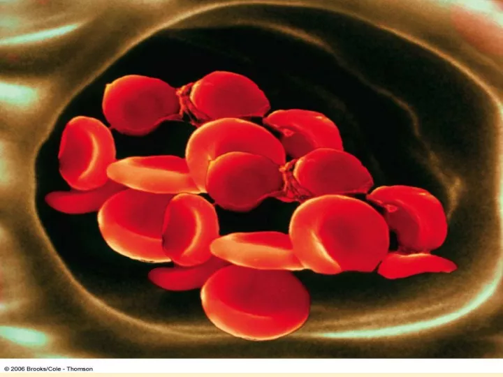
Protein Structure 1° structure: the sequence of amino acids in a polypeptide chain, read from the N-terminal end to the C-terminal end • Amino acid sequence (1° structure) of a protein determines its 3D structure which determines its properties and its biological function. • A striking example of the importance of primary structure is sickle-cell anemia, a disease caused by a change in one amino acid in each of two of the four chains of hemoglobin(HbS the β-chain of sickle cell Hb a valine residue has replaced a glutamic acid residue at position 6).
2° structure: the ordered 3-dimensional arrangements (conformations) in localized regions of a polypeptide chain; refers only to H-bonded arrangement of the peptide backbone e. g., -helix and -pleated sheet In Alzheimer’s disease patients, levels of β-amyloid become elevated, and this protein undergoes a conformational transformation from a soluble α helix–rich state to a state rich in β sheet and prone to self-aggregation.
-Helix Characteristics: • coil of the helix is clockwise or right-handed • there are 3.6 amino acids per turn • full turn distance is 5.4 Å (pitch) • each peptide bond is trans and planar • C=O of each peptide bond is hydrogen bonded to the N-H of the fourth amino acid away • C=O…...H-N hydrogen bonds are parallel to helical axis • all R groups point outward from helix • R groups are not involved in the H-bonds
-Pleated Sheet • polypeptide chains are folded back on itself • polypeptide chains lie adjacent to one another; may be parallel or antiparallel • R-groups alternate, first above and then below plane • each peptide bond is trans and planar • C=O and N-H groups of each peptide bond are perpendicular to axis of the sheet • C=O…...H-N hydrogen bonds are between adjacent sheets and perpendicular to the direction of the sheet
-Pleated Sheet antiparallel parallel • Reverse turn: allows peptide chain to reverse (bend) direction • Proline and glycine are prevalent
Tertiary (3°) structure: the 3D arrangement in space of all atoms in a polypeptide chain • Bonds stabilize the 3° structure: Metal ion coordination, side chain H-bond, electrostatic attraction, disulfide bond, and hydrophobic interaction • Quaternary (4°) structure: the association of polypeptide chains. • Proteins are divided into two large classes based on their three-dimensional structure. Protein Classifications according to shape : • fibrous proteins • globular proteins
Fibrous Proteins • Fibrous proteins: contain polypeptide chains organized approximately parallel along a singleaxis. They • consist of long fibers or large sheets • tend to be mechanically strong • are insoluble in water and dilute salt solutions • Play an important structural roles in nature
Collagen triple helix Collagen has an unusual amino acid composition and sequence: Glycine is found at almost every third residue Proline (Pro) makes up about 17% of collagen Collagen contains two uncommon derivative amino acids not directly inserted during translation. These amino acids are found at specific locations relative to glycine and are modified post-translationally by different enzymes, both of which require vitamin Cas a cofactor.
Hydroxyproline (Hyp), derived from proline. Hydroxylysine(Hyl), derived from lysine (Lys). Depending on the type of collagen, varying numbers of hydroxylysines are glycosylated (mostly having disaccharides attached). Cortisol stimulates degradation of (skin) collagen into amino acids.
The best-known defect in collagen biosynthesis is Scurvy, a result of a dietary deficiency of vitamin C required by prolyl and lysyl hydroxylases. • The resulting deficit in the number of hydroxyproline and hydroxylysine residues undermines the conformational stability of collagen fibers, leading to bleeding gums, swelling joints, poor wound healing, and ultimately to death. • Menkes’syndrome, characterized by kinky hair and growth retardation, reflects a dietary deficiency of the copper required by lysyl oxidase, which catalyzes a key step in formation of the covalent cross-links that strengthen collagen fibers. • ``
Genetic disorders of collagen biosynthesis include : • several forms of osteogenesis imperfecta, characterized by fragile bones. In Ehlers-Dahlos syndrome, a group of connective tissue disorders that involve impaired integrity of supporting structures, defects in the genes that encode α collagen-1, procollagen N-peptidase, or lysyl hydroxylase result in mobile joints and skin abnormalities.
Globular Proteins • Globular proteins: proteins which are folded to a more or less spherical shape • they tend to be soluble in water and salt solutions • most of their polar side chains are on the outside and interact with the aqueous environment by hydrogen bonding and ion-dipole interactions • most of their nonpolar side chains are buried inside • nearly all have substantial sections of -helix and -sheet • Example • Myoglobin
Myoglobin • Function in O2 storage in muscles • a single polypeptide chain of 153 amino acids • Compact with a single heme group in a hydrophobic pocket • 8 regions of -helix; no regions of -sheet • most polar side chains are on the surface • nonpolar side chains are folded to the interior • two His side chains are in the interior, involved with interaction with the heme group
Heme is a prosthetic group made from one protoporphyrin ring and iron in the center protoporphyrin ring is made from 4 pyrrole rings Fe(II) of heme has 6 coordinates sites; 4 sites interact with N atoms of protoporphyrin , 1 with N of a His side chain, and 1 with either an O2 molecule or an N of the second His side chain
Myoglobin • In the presence of globin, Fe(II) affinity to O2 increases and its affinity to CO decreases. • In the absence of globin, Fe(II) has high affinity to CO while low affinity to O2 . • In the absence of globin, Fe(II) can be easily oxidized to Fe(III) which has no affinity to O2
Globular Protein Subunits Alcohol dehydrogenase 2 Triosephosphate isomerase 2 Aldolase 3 Hemoglobin 2 + 2 Lactate dehydrogenase 4 Pyruvate kinase 4 2 Insulin Quaternary Structure • Quaternary (4°) structure: the association of polypepetide monomers into multisubunit proteins (dimer, trimer, tetramer, etc) • examples we will see in this course
Oxygen Binding of Hb • Hb is a tetramer of two -chains (141 amino acids each) and two -chains (153 amino acids each); a2b2 • each chain has 1 heme group; hemoglobin can bind up to 4 molecules of O2 • binding is cooperative; when one O2 is bound, it becomes easier for the next O2 to bind (positive cooperativity) • the function of hemoglobin is to transport oxygen • the structure of oxygenated Hb (loaded) is different from that of unoxygenated Hb (unloaded) • H+, CO2, Cl-, and 2,3-bisphosphoglycerate (BPG) affect the ability of Hb to bind and transport oxygen
Oxygen Binding of Hb O2 binding of hemoglobin and myoglobin hyperbolic sigmoidal
Oxygen Binding of Hb • The effect of pH on the oxygen-binding ability of Hb is called the Bohr effect • as pH decreases (more acidic), oxygen is released • Hb has lower affinity to O2 under [H+] • CO2 promotes release of O2 from HbO2
Oxygen Binding of Hb The Bohr effect
Actively Metabolizing Muscle Lungs + Higher pH than actively Lower pH due to production of H metabolizing tissue Hemoglobin binds O Hemoglobin releases O 2 2 + + Hemoglobin releases H Hemoglobin binds H Oxygen Binding of Hb Summary of the Bohr effect
Hemoglobin (Hb) • Hemoglobin in blood is bound to BPG • interaction is electrostatic, between negative charges on BPG and positive side chains (e.g., Lys, Arg) of hemoglobin • BPG promotes O2dissociation • Hb stripped of BPG remains saturated with O2
Fetal Hemoglobin, Hb F • has a higher affinity for O2 than maternal Hb A • structure is a2g2 • binds less strongly to BPG than does Hb A Oxygen binding capacity of Hb F
Adaptation to High Altitude • Physiologic changes that accompany prolonged exposure to high altitude include an increase in the number of erythrocytes and in their concentrations of hemoglobin and of BPG. • Elevated BPG lowers the affinity of HbA for O2 (decreases P50), which enhances release of O2 at the tissues.
BIOMEDICAL IMPLICATIONS Myoglobinuria • Following massive crush injury, myoglobin released from damaged muscle fibers colors the urine dark red. • Myoglobin can be detected in plasma following a myocardial infarction. Anemias • Anemias, reductions in the number of red blood cells or of hemoglobin in the blood, can reflect impaired synthesis of hemoglobin (eg, in iron deficiency; or impaired production of erythrocytes (eg, in folic acid or vit.B12 def.)
Glycosylated Hemoglobin (HbA1c) When blood glucose enters the erythrocytes it glycosylates the amino group of lysine residues and the amino terminals of hemoglobin. The fraction of hemoglobin glycosylated, normally about 5%, is proportionate to blood glucose concentration. Since the half-life of an erythrocyte is typically 60 days, the level of glycosylated hemoglobin (HbA1c) reflects the mean blood glucose conc.over the preceding 6–8 weeks. Measurement of HbA1c therefore provides valuable information for management of diabetes mellitus.
Hemoglobin (HbS) • In sickle cell, Val replaces the β6 Glu of HbB, creating a “sticky patch” that has a complement on deoxyHb (but not on oxyHb). • DeoxyHbS polymerizes at low O2 concentrations, forming fibers that distort erythrocytes into sickle shapes. • Alpha and beta thalassemias are anemias that result from reduced production of α and β subunits of HbA, respectively
Factors Directing Folding • Noncovalent interactions, such as • hydrogen bonding between polar side chains, e.g., Ser and Thr • hydrophobic interaction between nonpolar side chains, e.g., Val and Ile • electrostatic attraction between side chains of opposite charge, e.g., Lys and Glu • electrostatic repulsion between side chains of like charge, e.g., Lys and Arg, Glu and Asp • Formation of disulfide (-S-S-) covalent bonds between side chains of cysteines
Denaturation • Denaturation: the unfolding of the protein the loss of biological activity • Native protein (active) denatured protein (inactive) • Denaturation can be brought about by • Heat • Large changes in pH, which alter charges on side chains, e.g., -COO- to -COOH or -NH+ to -NH • Detergents such as sodium dodecyl sulfate (SDS) which disrupt hydrophobic interactions • Urea or guanidine HCL, which form H-bonds with protein that are stronger than those within the protein disrupt H-bonding • Mercaptoethanol, which reduces disulfide bonds.