Download

1 / 119
1.23k likes | 2.1k Views
Syphilis. David M. Bracciano, D.O. Syphilis. Aka lues Contagious, sexually transmitted disease Spirochete Treponema pallidum Enters through skin or mucous membrane where primary manifestations are seen. Treponema pallidum. Spiral spirochete that is mobile

E N D
Syphilis David M. Bracciano, D.O.
Syphilis • Aka lues • Contagious, sexually transmitted disease • Spirochete Treponema pallidum • Enters through skin or mucous membrane where primary manifestations are seen
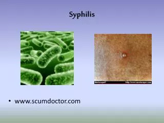
Treponema pallidum • Spiral spirochete that is mobile • # of spirals varies from 4 to 14 • Length 5 to 20 microns • Can be seen on fresh primary or secondary lesions by darkfield microscopy or fluorescent antibody techniques
Treponema pallidum • Motility has three movements • Projection and rotation in the direction of the long axis • Bending or twisting from side to side • Pathogenic in apes, humans, and rabbits
Syphilis epidemiology • Major health problem throughout world • U.S. 2.6 cases per 100,000 in 1999 • Lowest level ever recorded • Concentrated in 28 counties SE U.S. • Mainly gay men and crack cocaine users
Syphilis epidemiology • Enhances risk of transmission of HIV • HIV testing recommended in all patients with syphilis • Reportable disease, contact tracing
Serologic Tests • Reveal patients immune status not whether they are currently infected • Use lipoidal antigens rather than T. pallidum or components of it; non-treponemal antigen tests • RPR; rapid plasma reagin • VDRL; Venereal Disease Research Laboratory
Serologic Tests • Positive within 5 to 6 weeks after infection • Strongly positive in secondary phase • Strength of reaction is stated in dilutions • May become negative with treatment or over decades
Serologic Tests • To improve sensitivity and specificity tests using a specific treponemal antigen devised • MHA-TP: microhemagglutination assay for T. pallidum • FTA-ABS: fluorescent treponemal antibody absorption test • All positive nontreponemal test results should be confirmed with a specific treponemal test
Serologic Tests • Treponemal tests become positive early, useful in confirming primary syphilis • Remain positive for life, useful in diagnosing late disease • Treatment results in loss of positivity in 13-24% of patients
Biologic False-Positive Test Results • Positive STS in persons with no history or clinical evidence of syphilis • Acute BFP: those that revert to negative in less than 6 months • Chronic BFP: persist > 6 months
Acute BFP Vaccinations Infections pregnancy Chronic BFP Connective tissue disease (SLE) Liver disease Blood transfusions IVDA BFP Test Results in Syphilis
Cutaneous Syphilis • Chancre is usually the first cutaneous lesion • 18 to 21 days after infection • Round indurated papule with eroded surface that exudes a serous fluid • Cartilage-like consistency • Usually painless (Hunterian chancre is “classic” heals without scarring)
Chancre • Inguinal adenopathy 1-2 weeks after chancre • Generally occur singly, may be multiple • Diameter mm to cm
Chancres • Women genital chancre less often observed due to location within the vagina and cervix • Edema of labia may occur
The “Dory Flop” • “a chancre in the prepuce, being too hard to bend, will flip over all at once when the prepuce is drawn back” • Resembles the movement of a dory as it is being turned upside down
Chancre • Untreated, the chancre heals spontaneously in 1 to 4 months • Constitutional symptoms begin just as chancres disappear • Extragenital chancre: may be larger, frequently on lips, rarely tongue, tonsil, breast, finger, anus.
Chancre Histology • Ulcer covered by neutrophils and fibrin • Dense infiltrate of lymphocytes and and plasma cells • Spirochetes in untreated primary syphilis with silver stains; Warthin-Starry • Direct fluorescent antibody tissue test (DFAT-TP)- serous exudate collected on a slide sent for exam
Serology • Nontreponemal tests positive 50% • Treponemal tests positive 90% • Positivity depends upon duration of infection, if chancre has been present for several weeks, test is usually positive
Incubation 3 weeks Painless, no ulcer, no surrounding inflammatory zone Oval, hard Lymphadenopathy may be bilateral, nontender, nonsuppurative Incubation 4-7 days Ulcer inflamed, very painful, inflammatory zone Soft, covered by a membrane Lymphadenopathy unilateral, tender, suppurative Chancre vs. Chancroid
Ddx in Syphilis • Chancroid; multiple lesions, may coexist with chancre, must r/o syphilis • Granuloma Inguinale; indurated nodule that erodes, soft red granulation tissue, Donovan’s bodies in macrophages with Wright’s or Giemsa’s stain • Lymphogranuloma Venereum; small, painless, superficial non indurated ulcer, primary lesions followed in 7 to 30 days by adenopathy • HSV; grouped vesicles, burning pain
Secondary Syphilis • Skin manifestations in 80% called syphilids • Symmetric, generalized, superficial, macular transient; later papular, pustular • Early on face, shoulders, flanks, palms and soles, anal or genital areas
Secondary Syphilis Macular Eruptions • Exanthematic erythema 6-8 weeks after chancre, extends rapidly, may last hours to months • Round indistinct, slightly scaling ham-colored macules • Pain, burning absent, pruritus may be present • Generalized shotty adenopathy
Secondary Syphilis Papular Eruptions • Arise later than macular, raw-ham, round, 2-5mm or more in diameter, slightly raised, smooth or thick scale • Face and flexures of arms and legs, trunk • Palmar and plantar yellowish-red spots • Ollendorf’s sign; papule tender to touch of a blunt probe
Secondary Syphilis Papular Eruptions • Papulosquamous syphilids may produce a psoriasiform eruption • Follicular or lichenoid syphilids appear as minute scale-capped papules • Tend to be disseminated, but may be localized, asymmetrical, con figurate, hypertrophic, confluent.
Secondary SyphilisPapular Eruptions • Annular syphilid mimics sarcoidosis, more common in blacks • Cheeks, angle of mouth, annular, gyrate, ridges; “nickels and dimes” • Pustular syphilid; rare, face, trunk, extremities red small crust-covered ulceration • Rupial syphilid; superficial ulceration is covered with a pile of terraced crusts resembling an oyster shell.
Secondary SyphilisPapular Eruptions • Lues Maligna; rare, severe ulcerations, pustules, or rupioid lesions, accompanied by severe constitutional symptoms. • Condylomata lata; papular mass, weeping, gray 1-3cm, groin, anus (not vegetative like condylomata acuminata) • Syphilitic alopecia; irregular, scalp has a moth-eaten appearance 5% of pts
Secondary SyphilisMucous Membrane • Present in 1/3 of secondary syphilis • Most common is “syphilitic sore throat” • Diffuse pharyngitis, hoarseness • Tongue; patches of desquamation of papillae • Ulcerations of tongue and lips in late stages
Secondary SyphilisMucous membrane • Mucous patches are the most characteristic mucous membrane lesions; macerated, flat. Grayish, rounded erosions covered by a delicate, soggy membrane. • Highly infectious, occur on tonsils, tongue, pharynx, gums, lips, and buccal areas, or on the genitalia