Download
1 / 37
490 likes | 1.52k Views
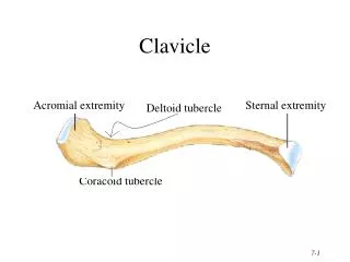
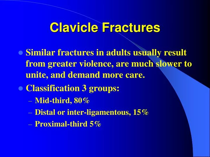
Clavicle Fractures. Similar fractures in adults usually result from greater violence, are much slower to unite, and demand more care. Classification 3 groups: Mid-third, 80% Distal or inter-ligamentous, 15% Proximal-third 5%. Clavicle Fractures.

E N D
Clavicle Fractures • Similar fractures in adults usually result from greater violence, are much slower to unite, and demand more care. • Classification 3 groups: • Mid-third, 80% • Distal or inter-ligamentous, 15% • Proximal-third 5%
Clavicle Fractures • Among the most common fracture occurring in children. • In children usually heal without problems (the saying goes that if you put two ends of a fractured clavicle in the same room [pediatric] they will heal).
Clavicle Fractures • Ideal method of management has not yet been developed. • Over 200 methods have been described. • Most do well with nonoperative management. • It may take at least 3 months for adults to resume heavy activities.
Clavicle Fractures Distal clavicle fractures more problematic if involving the coracoclavicular ligaments. fracture without wts. with wts.
Clavicle Fractures Distal clavicle plate
Clavicle Fractures ORIF Displaced
Clavicle Fractures IM Rod
AC Separations • MOI- direct force that occurs from a fall on the point of the shoulder. • Major deformity is the downward displacement of the shoulder.
AC Separations • Classification:6 types of separation. • Types I-III most common. • Grade I - mild forces, Grade 6 - occurs with major forces.
AC Separation • Stress X-rays to differentiate between Grade I and Grade 3. • Gr. III has upward displacement 25-100% compared to the normal.
Grade I-III is usually conservative. Sling for comfort. Early ROM. Grade IV-VI is usually surgical. Grade II injuries can develop DJD. Grade III injuries can be repaired in a young laborer who performs overhead work. AC Separation Treatment
The Elbow • Little Leaguer’s elbow • Osteochondritis dissicans of the capitellum • Panner’s disease-osteochondrosis of the capitellum
Little Leaguer’s Elbow • A term used to describe a number of overuse conditions about the elbow associated with repetitive throwing that affects the skeletally immature elbow • MOI is valgus extension overload, which leads to traction stress on the medial aspect of the elbow, the medial collateral ligament, and the medial epicondyle
Little Leaguer’s Elbow • Valgus extension overload also results in compression stresses on the lateral aspect of the joint, leading to osteochondritis dissicans of the capitellum, loose bodies, and radial head overgrowth • The extension component can cause repetitive irritation of the olecranon in the olecranon fossa, which can lead to impingement & loose bodies
Little Leaguer’s Elbow Symptoms • Medial pain • Diminished throwing effectiveness • Decreased throwing distance
Little Leaguer’s Elbow Examination • Tenderness • Swelling over medial epicondyle • Elbow flexion contraction > 15 degrees • X-ray- fragmentation and widening of the epiphyseal lines
Little Leaguer’s Elbow Treatment • Rest ( 4-6 weeks) • No throwing • Ice • NSAID’s • May strengthen when pain free (>6wk)
Osteochondritris Dissicans Of The Humeral Capitellum • Represents an island of subchondral bone and its articular cartilage that begins to separate from the rest of the humerus • Symptoms include lateral pain, which is dull and worsens with motion, which locks and catches
Osteochondritris Dissicans Of The Humeral Capitellum • Etiology is unclear • Repetitive stress most likely cause • May have genetic predisposition • Between ages 10-15 • Common in throwers and gymnasts
Osteochondritris Dissicans Of The Humeral Capitellum Examination • Radiocapitellar tenderness • Flexion contracture • Crepitation • Effusion
Osteochondritris Dissicans Of The Humeral Capitellum Radiology • Crescent shaped region of sclerotic subchondral bone at the humeral capitellum • Possible loose bodies
Osteochondritris Dissicans Of The Humeral Capitellum Treatment • If no evidence of separation then rest, ice, ROM, and analgesics • Repeat X-ray check for healing • Surgery if locking, loose bodies, fragment separation or failure of conservative management
Panner’s Disease • Osteochondrosis of the humeral capitellum • Repetitive valgus stress causes compressive stress across the radiocapitellar • Occurs between 7-12 years of age ( peak incidence at 9 years) • May be susceptible at this time due to limited blood supply
Panner’s Disease Pathophysiology • Unknown • May be similar to Legg-Calve`-Perthes disease
Panner’s Disease Symptoms • Fairly sudden pain • Deep and dull achiness • Worsened with throwing • No mechanical symptoms (locking or catching)
Panner’s Disease Physical exam • Tenderness and swelling over the lateral elbow • Mild to moderate flexion contractures (usually from 5-20 degrees)
Panner’s Disease Radiology Fragmentation of the capitellum, with alternating area of sclerosis and rarefaction and an irregular joint surface
Panner’s Disease Treatment • Conservative • Complete rest from throwing until symptoms subside and ROM is normal • Repeat X-rays to monitor remodeling • May return when X-rays and exam is normal • Long term disability is rare
The Wrist Gymnast wrist Torus fractures Wrist fractures
Gymnast Wrist • Chronic overuse injury occurring at the physis if skeletally immature gymnasts • Presents with wrist pain • Usually due to repetitive hyperextension and overuse • Arms are used as weight bearing devices • Salter-Harris type I injury
Gymnast Wrist • Gymnast’s wrist frequently show physeal irregularities and bony sclerosis on X-ray • If untreated can result in permanent radial deformity and shortening due to growth arrest • Rest relieves symptoms • Extension splints can prevent recurrence