Download
1 / 28
280 likes | 364 Views
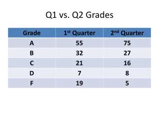
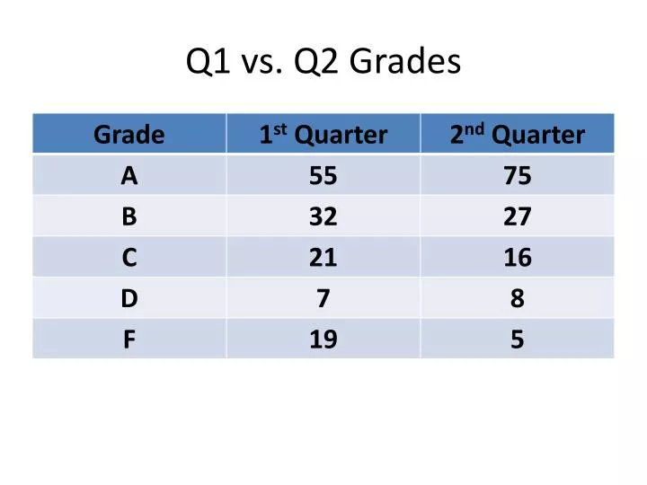
Q1 vs. Q2 Grades. Attachment. The T4 phage uses its tail fibers to bind to specific receptor sites on the outer surface of an E. coli cell.

E N D
Attachment. The T4 phage usesits tail fibers to bind to specificreceptor sites on the outer surface of an E. coli cell. Entry of phage DNA and degradation of host DNA.The sheath of the tail contracts,injecting the phage DNA intothe cell and leaving an emptycapsid outside. The cell’sDNA is hydrolyzed. 1 2 4 3 5 Release. The phage directs productionof an enzyme that damages the bacterialcell wall, allowing fluid to enter. The cellswells and finally bursts, releasing 100 to 200 phage particles. Phage assembly Synthesis of viral genomes and proteins. The phage DNAdirects production of phageproteins and copies of the phagegenome by host enzymes, usingcomponents within the cell. Assembly. Three separate sets of proteinsself-assemble to form phage heads, tails,and tail fibers. The phage genome ispackaged inside the capsid as the head forms. Head Tail fibers Tails Figure 18.6 The lytic cycle of phage T4, a virulent phage
Phage DNA The phage attaches to a host cell and injects its DNA. Many cell divisions produce a large population of bacteria infected with the prophage. Phage DNA circularizes Phage Occasionally, a prophage exits the bacterial chromosome, initiating a lytic cycle. Bacterial chromosome Lytic cycle Lysogenic cycle Certain factors determine whether The bacterium reproduces normally, copying the prophage and transmitting it to daughter cells. The cell lyses, releasing phages. Prophage Lytic cycle is induced Lysogenic cycle is entered or New phage DNA and proteins are synthesized and assembled into phages. Phage DNA integrates into the bacterial chromosome,becoming a prophage. Figure 18.7 The lytic and lysogenic cycles of phage , a temperate phage
The virus fuses with the cell’s plasma membrane. The capsid proteins are removed, releasing the viral proteins and RNA. 1 HIV Membrane of white blood cell 2 Reverse transcriptase catalyzes the synthesis of a DNA strand complementary to the viral RNA. HOST CELL 3 Reverse transcriptase catalyzes the synthesis ofa second DNA strand complementary to the first. Reverse transcriptase Viral RNA RNA-DNAhybrid 4 The double-stranded DNA is incorporated as a provirus into the cell’s DNA. 0.25 µm HIV entering a cell DNA NUCLEUS Provirus ChromosomalDNA RNA genomefor the nextviral generation 5 Proviral genes are transcribed into RNA molecules, which serve as genomes for the next viral generation and as mRNAs for translation into viral proteins. mRNA 6 The viral proteins include capsid proteins and reverse transcriptase (made in the cytosol) and envelope glycoproteins (made in the ER). 7 Capsids are assembled around viral genomes and reverse transcriptase molecules. 8 Vesicles transport the glycoproteins from the ER to the cell’s plasma membrane. 9 New viruses bud off from the host cell. New HIV leaving a cell Figure 18.10 The reproductive cycle of HIV, a retrovirus
The Genetics of Viruses & Bacteria • What do you know about viruses? • How big are viruses? • What are the components of a virus? • How do viruses identify appropriate cells to infect? • What is the lytic cycle of a bacteriophage? • What is the lysogenic cycle of a bacteriophage? • How do retroviruses (like HIV) reproduce? • How do “new” viruses emerge? • Mutation of an existing virus since there is no proofreading • Spread of an existing virus from 1 host species to another • Spread of viral disease from a small isolated population • 9. What is the difference between horizontal & vertical transmission? • Horizontal – 1 organism spreads to another • Vertical – 1 organism inherits disease from parent • 10. What are viroids & prions? • Viroids – tiny molecules of naked, circular RNA that infect plants, • several hundred nucleotides long • Prions – infectious proteins (NO genetic material) • Slow incubation period – at least 10 yrs • Virtually indestructible • 1997 Nobel Prize in Medicine – Stanley Prusiner
The Genetics of Viruses & Bacteria • What do you know about viruses? • How big are viruses? • What are the components of a virus? • How do viruses identify appropriate cells to infect? • What is the lytic cycle of a bacteriophage? • What is the lysogenic cycle of a bacteriophage? • How do retroviruses (like HIV) reproduce? • How do “new” viruses emerge? • 9. What is the difference between horizontal & vertical transmission? • 10. What are viroids & prions? • 11. How is bacterial DNA different from eukaryotic DNA? (refer to Ch. 19 notes) • Bacterial Eukaryotic • Circular chromosome Linear chromosomes • Nucleoid region Nucleus • No introns (all exons) Introns & exons • Transcription coupled w/ translation Transcription & translation separate • More mutations Fewer mutations (proofreading) • How does bacterial DNA replicate its circular chromosome? • - Figure 16.16
Phage DNA B+ Phage infects bacterial cell that has alleles A+ and B+ A+ 1 Host DNA (brown) is fragmented, and phage DNA and proteins are made. This is the donor cell. A+ 2 B+ Donorcell A bacterial DNA fragment (in this case a fragment withthe A+ allele) may be packaged in a phage capsid. 3 A+ Crossingover Phage with the A+ allele from the donor cell infects a recipient A–B– cell, and crossing over (recombination) between donor DNA (brown) and recipient DNA (green) occurs at two places (dotted lines). 4 A+ A– B– Recipientcell The genotype of the resulting recombinant cell (A+B–) differs from the genotypes of both the donor (A+B+) and the recipient (A–B–). 5 A+ B– Recombinant cell Figure 18.16 Generalized transduction
1 m Sex pilus Figure 18.17 Bacterial conjugation
F Plasmid Bacterial chromosome F+ cell F+ cell Mating bridge F+ cell Bacterial chromosome F– cell A cell carrying an F plasmid(an F+ cell) can form amating bridge with an F– celland transfer its F plasmid. DNA replication occurs inboth donor and recipientcells, using the single parental strands of the F plasmid as templates to synthesize complementary strands. The plasmid in the recipient cell circularizes. Transfer and replication result in a compete F plasmid in each cell. Thus, both cells are now F+. A single strand of the F plasmid breaks at a specific point (tip of blue arrowhead) and begins tomove into the recipient cell. As transfer continues, the donor plasmid rotates(red arrow). 3 2 6 4 1 8 4 3 2 1 7 5 (a) Conjugation and transfer of an F plasmid from an F+ donor to an F– recipient Hfr cell F+ cell F factor The circular F plasmid in an F+ cellcan be integrated into the circularchromosome by a single crossoverevent (dotted line). The resulting cell is called an Hfr cell (for High frequency of recombination). B+ D+ C+ C+ A+ D+ A+ A+ B+ D+ D+ A+ C+ C+ B+ B+ A+ B+ C– C– C– C– F– cell B– B+ D– D– D– A+ B– B– D– B– A– A– A– A– A+ Since an Hfr cell has all the F-factor genes, it can form a mating bridge with an F– cell and transfer DNA. The location and orientation of the F factor in the donor chromosome determine the sequence of gene transfer during conjugation. In this example, the transfer sequence for four genes is A-B-C-D. A single strand of the F factorbreaks and begins to move through the bridge. DNA replication occurs in both donor and recipient cells, resulting in double-stranded DNA The mating bridgeusually breaks well before the entire chromosome and the rest of the F factor are transferred. (b) Conjugation and transfer of part of the bacterial chromosome from an Hfr donor to an F– recipient, resulting in recombination Temporary partial diploid C– C– Recombinant F– bacterium B+ B– D– D– B+ B– A– A– A+ A+ The piece of DNA ending up outside thebacterial chromosome will eventually be degraded by the cell’s enzymes. The recipient cell now contains a new combination of genes but no F factor; it is a recombinant F– cell. Two crossovers can result in the exchange of similar (homologous) genes between the transferred chromosome fragment (brown) and the recipient cell’s chromosome (green). Figure 18.18 Conjugation and recombination in E. coli Plasmid – extra-chromosomal, small, circular, self-replicating DNA
trp operon Promoter Promoter Genes of operon RNA polymerase Start codon Stop codon trpR trpD trpC trpB trpE trpA DNA Operator Regulatory gene 3 mRNA 5 mRNA 5 C E D B A Polypeptides that make up enzymes for tryptophan synthesis Inactiverepressor Protein (a) Tryptophan absent, repressor inactive, operon on. RNA polymerase attaches to the DNA at the promoter and transcribes the operon’s genes. Figure 18.21 The trp operon: regulated synthesis of repressible enzymes
DNA No RNA made mRNA Active repressor Protein Tryptophan (corepressor) (b) Tryptophan present, repressor active, operon off. As tryptophan accumulates, it inhibits its own production by activating the repressor protein.
Promoter Regulatorygene Operator DNA lacl lacZ NoRNAmade 3 RNApolymerase mRNA 5 Activerepressor Protein (a) Lactose absent, repressor active, operon off. The lac repressor is innately active, and inthe absence of lactose it switches off the operon by binding to the operator. Figure 18.22 The lac operon: regulated synthesis of inducible enzymes
lac operon DNA lacl lacz lacY lacA RNApolymerase 3 mRNA 5 mRNA 5' mRNA 5 -Galactosidase Permease Transacetylase Protein Inactiverepressor Allolactose(inducer) (b) Lactose present, repressor inactive, operon on. Allolactose, an isomer of lactose, derepresses the operon by inactivating the repressor. In this way, the enzymes for lactose utilization are induced.
Before the invention of antibiotics, the clean modern hospitals of India, which were mostly reserved for Europeans, reported cholera death rates of 86%. Meanwhile, in other more crowded and less hygienic hospitals, the death rate from cholera was only 27%. WHY? Based on knowledge gained from the case study, explain the most likely process that occurred in the crowded hospitals that led to such a low death rate from cholera.
(a) Animal development. Most animals go through some variation of the blastula and gastrula stages. The blastula is a sphere of cells surrounding a fluid-filled cavity. The gastrula forms when a region of the blastula folds inward, creating a tube—a rudimentary gut. Once the animal is mature, differentiation occurs in only a limited way—for the replacement of damaged or lost cells. Gut Cell movement Zygote (fertilized egg) Eight cells Blastula (cross section) Gastrula (cross section) Adult animal (sea star) Cell division Morphogenesis Observable cell differentiation (b) Plant development. In plants with seeds, a complete embryo develops within the seed. Morphogenesis, which involves cell division and cell wall expansion rather than cell or tissue movement, occurs throughout the plant’s lifetime. Apical meristems (purple) continuously arise and develop into the various plant organs as the plant grows to an indeterminate size. Seed leaves Shoot apical meristem Root apical meristem Zygote (fertilized egg) Two cells Embryo inside seed Plant Figure 21.4a, b
Chapter 21: The Genetic Basis of Development • How do we study development in the genetics-based lab? • How does a zygote transform into an organism? • How do cells become differentiated? -All cells have the same DNA, so differential gene expression must be the explanation!
Egg cell donor Mammary cell donor APPLICATION This method is used to produce cloned animals whose nuclear genes are identical to the donor animal supplying the nucleus. 1 2 Egg cell from ovary Nucleus removed Nucleus removed Cells fused Cultured mammary cells are semistarved, arresting the cell cycle and causing dedifferentiation 3 TECHNIQUE Shown here is the procedure used to produce Dolly, the first reported case of a mammal cloned using the nucleus of a differentiated cell. Nucleus from mammary cell Grown in culture 4 RESULTS The cloned animal is identical in appearance and genetic makeup to the donor animal supplying the nucleus, but differs from the egg cell donor and surrogate mother. Early embryo Implanted in uterus of a third sheep 5 Surrogate mother Embryonic development 6 Lamb (“Dolly”) genetically identical to mammary cell donor Figure 21.7
Chapter 21: The Genetic Basis of Development Embryonic stem cells Adult stem cells Early human embryo at blastocyst stage (mammalian equiva- lent of blastula) From bone marrow in this example Totipotent cells Pluripotent cells Cultured stem cells Different culture conditions Liver cells Blood cells Nerve cells Different types of differentiated cells Figure 21.9 4. What is a stem cell? -a relatively unspecialized cell -can differentiate into cells of different types under specific conditions -Embryonic = totipotent -Adult = pluripotent (can produce some, but not all, cell types)
Chapter 21: The Genetic Basis of Development 2 1 Nucleus Master control gene myoD Other muscle-specific genes DNA OFF OFF Embryonicprecursor cell Determination. Signals from othercells lead to activation of a masterregulatory gene called myoD, andthe cell makes MyoD protein, atranscription factor. The cell, nowcalled a myoblast, is irreversiblycommitted to becoming a skeletalmuscle cell. OFF mRNA MyoD protein(transcriptionfactor) Myoblast (determined) Differentiation. MyoD protein stimulatesthe myoD gene further, and activatesgenes encoding other muscle-specifictranscription factors, which in turn activate genes for muscle proteins. MyoD also turns on genes that block the cell cycle, thus stopping cell division. The nondividing myoblasts fuse to become mature multinucleate muscle cells, alsocalled muscle fibers. mRNA mRNA mRNA mRNA Myosin, othermuscle proteins,and cell-cycleblocking proteins MyoD Anothertranscriptionfactor Muscle cell(fully differentiated) 5. What type of genetic signal leads to cell differentiation? -Step 1: Cell receives signals from other cells -Step 2: A regulatory gene is turned “on”, and a protein is made that activates other genes. (“point of no return”) -Step 3: Activated genes make proteins that determine cell type/ structure/behavior.
Epidermis Signal protein Gonad Anchor cell Vulval precursor cells Outer vulva ADULT Inner vulva Epidermis Figure 21.16b (b) Induction of vulval cell types during larval development.
EXPERIMENT RESULTS CONCLUSION Spemann and Mangold transplanted a piece of the dorsal lip of a pigmented newt gastrula to the ventral side of the early gastrula of a nonpigmented newt. Pigmented gastrula (donor embryo) Dorsal lip of blastopore Nonpigmented gastrula (recipient embryo) During subsequent development, the recipient embryo formed a second notochord and neural tube in the region of the transplant, and eventually most of a second embryo. Examination of the interior of the double embryo revealed that the secondary structures were formed in part from host tissue. Primary embryo Primary structures: Secondary (induced) embryo Secondary structures: Neural tube Notochord Notochord (pigmented cells) Neural tube (mostly nonpigmented cells) The transplanted dorsal lip was able to induce cells in a different region of the recipient to form structures different from their normal fate. In effect, the dorsal lip “organized” the later development of an entire embryo. Figure 47.25
Chapter 21: The Genetic Basis of Development Unfertilized egg cell Molecules of a a cytoplasmic determinant Fertilization Nucleus Zygote (fertilized egg) Mitotic cell division Two-celled embryo (a) Cytoplasmic determinants in the egg. The unfertilized egg cell has molecules in its cytoplasm, encoded by the mother’s genes, that influence development. Many of these cytoplasmic determinants, like the two shown here, are unevenly distributed in the egg. After fertilization and mitotic division, the cell nuclei of the embryo are exposed to different sets of cytoplasmic determinants and, as a result, express different genes. Figure 21.11a -cytoplasmic determinants in the unfertilized egg regulate gene expression in the zygote that affects differentiation/development
Chapter 21: The Genetic Basis of Development Tail Head T1 A8 T2 A7 T3 A6 A1 A5 A2 A4 A3 Wild-type larva Tail Tail A8 A8 A7 A6 A7 Mutant larva (bicoid) (a) Drosophila larvae with wild-type and bicoid mutant phenotypes. A mutation in the mother’s bicoid gene leads to tail structures at both ends (bottom larva). The numbers refer to the thoracic and abdominal segments that are present. Figure 21.14a -Cytoplasmic determinants from mother’s egg initially establish the axes of the body of Drosophila. -bicoid gene
Egg cell Nurse cells Developing egg cell 1 bicoid mRNA Bicoid mRNA in mature unfertilized egg 2 Fertilization 100 µm Translation of bicoid mRNA Bicoid protein in early embryo 3 Anterior end (b) Gradients of bicoid mRNA and Bicoid protein in normal egg and early embryo. Figure 21.14b
Chapter 21: The Genetic Basis of Development Hierarchy of Gene Activity in Early Drosophila Development Maternal effect genes (egg-polarity genes) Gap genes Segmentation genes of the embryo Pair-rule genes Segment polarity genes Homeotic genes of the embryo Other genes of the embryo -7. How does morphogenesis (pattern formation) occur in animals? After the body’s axes are determined (by cytoplasmic determinants)… -Segmentation genes produce proteins that direct formation of body segments. -Then, the development of specific features of the body segments is directed by HOMEOTIC GENES (Hox genes.)
Chapter 21: The Genetic Basis of Development Adult fruit fly Fruit fly embryo (10 hours) Fly chromosome Mouse chromosomes Mouse embryo (12 days) Adult mouse Figure 21.23 8. What is the relationship among the genetic basis of development across organisms? -Molecular analysis of the homeotic genes in Drosophila has shown that they all include a sequence called a homeobox -An identical (or very similar) DNA sequence has been discovered in the homeotic genes of vertebrates and invertebrates
Chapter 21: The Genetic Basis of Development Ced-9 protein (active) inhibits Ced-4 activity Mitochondrion Ced-4 Ced-3 Death signal receptor Inactive proteins Cell forms blebs (a) No death signal Ced-9 (inactive) Death signal Active Ced-3 Active Ced-4 Other proteases Nucleases Activation cascade (b) Death signal 9. What is apoptosis? -programmed cell death (cell suicide) Figure 21.18 Molecular basis of apoptosis in C. elegans
Chapter 21: The Genetic Basis of Development Interdigital tissue 1 mm Figure 21.19 8. What is apoptosis? -programmed cell death (cell suicide) -necessary for development of hands/feet in vertebrates