Download
1 / 12
150 likes | 783 Views
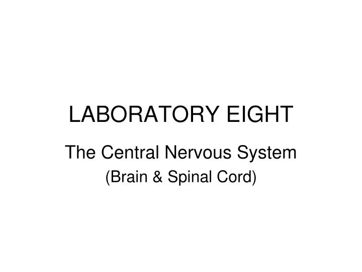
LABORATORY EIGHT. The Central Nervous System (Brain & Spinal Cord). Nervous Tissue. Neurons: functional cells that transport electrical impulses Neuroglia: non-conductive cells Schwann cells . Human Brain. Transverse fissure. Human Brain – Right Half. Septum pellucidum. Fornix.

E N D
LABORATORY EIGHT The Central Nervous System (Brain & Spinal Cord)
Nervous Tissue • Neurons: functional cells that transport electrical impulses • Neuroglia: non-conductive cells • Schwann cells
Human Brain Transverse fissure
Human Brain – Right Half Septum pellucidum Fornix Choroid Plexus Optic Chiasma Corpora quadrigemina Mammilary body Cerebral aqueduct Fourth ventricle
Human Brain Ventricles Site of massa intermedia
Cerebral Meninges(Three layers of protective connective Tissue in CNS)
Dura Mater in Cerebral Meninges(Fig 8.15, p140) • Modified in two areas: • Falx cerebri – penetrates longitudinal fissurebetween brain hemispheres • Tentorium cerebelli – penetrates transverse fissure that separates the cerebrum from the cerebellum
Cross-Section of Spinal Cord(Also study fig 8.12, p138) Dorsal gray horn White matter Ventral gray horn Central Canal
Electroencephalogram • EEG is a record of the electrical activity that takes place in the brain • Brain waves are characterized by their frequency (cycle per second) - Fig 8.16, p140 • Alpha (8-13 cps): relaxed with eyes closed • Beta (14-25 cps): alert, performing a task • Theta (4-7 cps): normal in children, but abnormal in adult - indicates emotional/mental imbalances • Delta (<4 cps): deep sleep • Report 8C, p149
Laboratory Report • P145, Q1 & P146, Q2 - bottom of the page • On Fig 8.3 and Fig 8.5, draw a thick black line that crosses thalamus • Table 8.1 • Compare the size of characteristics in question to the rest of the brain of the same species; e.g., the size of the cerebellum of human compare to the rest of the human brain
Sheep Brain DissectionGoogle search under “image” for Sheep brain dissection • Each lab pair should obtain a sheep brain, dissection tools, and a tray • Choose a sheep brain with intact pituitary gland • Note the 1800 relationship between the cerebrum, cerebellum, and spinal cord • Identify the superficial structures listed under 8B Procedure, step 1 (p134) and step 3 (p136) • Identify the longitudinal fissure and transverse fissure, but the central and lateral fissures can not be identified on the sheep brain • Grasp the sheep brain gently by the cerebral hemispheres and the cerebellum, and separate them at the transverse fissure to see the corpora quadrigemina (superior & inferior colliculi) & pineal body (step 5, p137) • To prepare for pituitary gland removal, carefully cut Trigeminal V cranial nerve, 1cm above its attachment site to the brain • When removing the pituitary gland, look underneath it to make sure no cranial nerve is attached • If you see a string structure attached to the pituitary gland from one side and to the floor of the brain from another side, clip it with a pair of scissors closer to the pituitary gland • Identify cranial nerves I, II, III, IV, V, VI and XI • Section the sheep brain by placing it ventral side up. • Make a long, smooth, midsagittal cut. Be sure to completely divide the brain in half • Identify the structures listed under step 2, p135 • Observe the prepared coronal section of the sheep brain and identify the assigned structure on step 7, p137 • Take half of the dissected brain home, and return them to the lab when done.