Download
1 / 18
181 likes | 575 Views
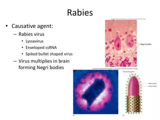
Rabies. Rabies is a viral infection of the central and peripheral NS ,that causes encephalitis with or without paralysis. Zoonosis. Family: Rhabdoviridae Genus : Lyssavirus Bullet shaped virion 75X 180 nm Envelop contains 10-nm spike-like gp G (=400 spike) Protein matrix

E N D
Rabies is a viral infection of the central and peripheral NS ,that causes encephalitis with or without paralysis. • Zoonosis Family: Rhabdoviridae Genus : Lyssavirus Bullet shaped virion 75X 180 nm Envelop contains 10-nm spike-like gp G (=400 spike) Protein matrix Helical nucleocapsid Nonsegmented ssRNA of –ve sense
The rabies virus genome is RNA of approximately 12 kb. There is a leader-sequence (LDR) of approximately 50 nucleotides, followed by N, P, M, G, and L genes.
The viruses are not viable outside of hosts and are inactivated by sunlight, heat, and desiccation. Within the host, the rabies viruses are highly neurotropic and replicate slowly within muscle cells
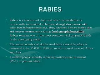
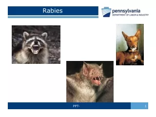
Transmission • Zoonosis • Dogs, followed by cats and other wild animals e.g. foxes • Bats always considered rabid. • Mode of transmission: • Inoculation of infected saliva through a rabid animal bite • Licking of animal on non intact skin • Transplantation : rare
Pathology • No gross abnormalities • Perivascular cuffing & cell degradation. • Negri’s bodies : pathognomic , eosinophilicintracytoplasmic inclusions (viral nucleocapsid aggregates)
4 clinical stages: • Incubation period: • Asymptomatic. • 1 – 3 m. (5 days -7 years; In rare cases, human rabies with an extended IP (2-7 y) has been reported). • Duration depends on: • Size of saliva inoculation • Size and depth of bite • Proximity of the brain. (age) • Prodromal period: • Nonspecific symptoms. • Pathognomicspesific symptoms
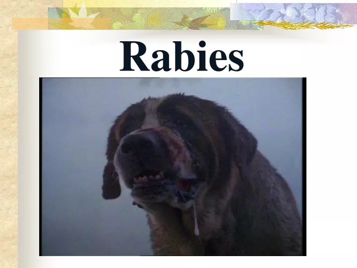
Neurologic stage : • Encephalitis or meningo-encephalitis • Two forms: • Furious form (encephalitic): • Hyperexcitability, Hyperactivity, and convulsions. • Hallucinations • Excessive salivation • Hydrophobia(abnormal contraction of diaphragm, respiratory, laryngeal, pharyngeal) • Aerophobia sometimes. • Dumb form (paralytic): • Generalized flaccid paralysis • Combined form
Coma, and is associated with MOF regardless of the form of presentation. • Hematemesis occurs in approximately 30–60% of patients during the last few hours of life. • Cardiac arrhythmias occur in almost all cases, with the cause of death related to cardiac and circulatory insufficiency. • It takes 4-16 days from the beginnig of the illness up to death. • Very few cases are reported to recover(~3)
Management: • Postexposureprohylaxis: • Wound care: • Wash with water, soap. Don’t scrub . Or • detergent (70% alcohol or ticture of aq. iodine). • Don’t suture unless excessive. • 2. Anti-tetanus • 3. Prophylactic antibiotics if indicated • 4. Immunoprophylaxis: both active & passive.
Passive immunization: • RIG, 20 IU/ml • Around the wound + I.M • Active immunization: • The following are safe and immunogenic: • Human cell diploid vaccine (HCDV) IM , on 0, 3,7,14,21, ±28 day • Rabies vaccine adsorbed (RVA) • Purified Chick embryo cell vaccine (PCEC) • Vero cell vaccine • Neural tissue vaccines are used in some developing countries: • simple vaccine: brain tissue vaccine, (infected brain +βpropol) . 2cc IM for 10 days. • in the past : 5 ml around umblicus( SE: allergic encephalomyelitis 1:2000- 4000) • B. Suckling mice brain tissue.
Pre-exposure prophylaxis: • For persons at risk: vet, lab workers, health officers • Travellers to endemic area. • 3 doses • Vaccination is effective for 2 years.
Antemortem diagnosis: • Ag detection: • Could be detected early • Skin biopsy: nape of the neck • Corneal impression: IF or ELISA • Virus isolation: • saliva or CSF • Cell culture or IT inoculation of mice. • RT-PCR • Ab detection: • Appears after 8 days • In serum : diagnostic in un vaccinated pts • In CSF is diagnostic ALWAYS. They appear after serum. Postmortem diagnosis: Brain biopsy: Sections from hippocampus, cerebellum….. Histopathology /IHC
Histopathologic evidence of rabies encephalomyelitis includes the following: • Mononuclear infiltration • Perivascular cuffing • Lymphocytic foci • Babes nodules consisting of glial cells • Negri bodies Babes Nodules Perivascular cuffing
Negri Enlargement of a Negri body in Sellers stained brain tissue. Note the basophilic (dark blue granules in the inclusion).
Rabies-infected neuronal cell with intracytoplasmic inclusions. The red stain indicates areas of rabies viral antigen by using IHC.
Rabies/Differential Diagnosis • Meningitis/Encephalitis: Japanese, eastern equine, West Nile V., enterovirus 71, • Epilepsy • Drug toxicity • Acute hepatic porphyria, neuropsychiatric disturbances • Substance abuse, acute serotonin syndrome • Pseudohydrophobia (hysterical reaction to animal bites)