Download
1 / 14
140 likes | 297 Views
Perception Chapter 3. Light is necessary but not sufficient for vision Ganzfeld : a visual field completely lacking in contour, or luminance changes. Prolonged exposure can cause perceptual blank out ( snow blindness).

E N D
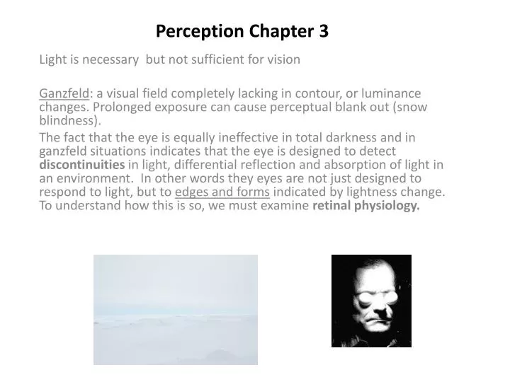
Perception Chapter 3 Light is necessary but not sufficient for vision Ganzfeld: a visual field completely lacking in contour, or luminance changes. Prolonged exposure can cause perceptual blank out (snow blindness). The fact that the eye is equally ineffective in total darkness and in ganzfeld situations indicates that the eye is designed to detect discontinuities in light, differential reflection and absorption of light in an environment. In other words they eyes are not just designed to respond to light, but to edges and forms indicated by lightness change. To understand how this is so, we must examine retinal physiology.
Retinal Physiology: a) the photoreceptors (rods and cones) are not the only neurons found in the retina. They synaps to collector cells, which in turn synapse to retinal ganglioncells b) While there are over 130 million photoreceptors, there are only about 1 million retinal ganglion cells. This means that RGC must summate across a number of photoreceptors c) Single cell recordings of RGC activity indicate that RGC do this by virtue of a receptive field organization.
Recording retinal ganglion cell activity Spot of light on one part of retina causes cell to increase firing rate (a: “on” response); another spot causes decrease (c: “off response). Same cell, just slight change in position of light. Note: B represents a “change” response.
RGC on/off receptive field organization Top: On center (white) off surround (grey) Bottom: off center (grey) on surround (white)
RGC on/off receptive field organization Receptive field: a limited spatial extend within the retina in which the activity of a RGC can be influenced. Lateral inhibition: the fact that stimulated adjacent regions tend to antagonistically effect one another. The receptive field organization of RGC acts to enhance edges or borders of adjacent regions of varying lightness, hence the outline of a form is exaggerated. Size of receptive fields tends to increase with increased retinal eccentricity. This indicates the macular dedication to resolution, or the ability to respond to small details of visual pattern vs. the peripheral dedication to sensitivity, or the ability to simply detect the presence of a lightness change. further evidence for this division of labor is found in the variations in the degree of convergence. In the fovea, photoreceptors and RGC have a nearly one to one correspondence. However, in the periphery, the degree of convergence can be several hundred to one. In fovea, sensitivity is sacrificed for resolution, in periphery resolution is sacrificed for sensitivity.
Retinal circuitry • Horizontal cells: amplify photoreceptor response • Bipolar cells: combine and summate photoreceptor response • Amacrine cells: Influence temporal pattern of RCG response
Types of RGC M Cells: generally larger, with larger receptive fields, more sensitive to small lightness differences within the field, but no sensitivity to wavelength, 80% of pop. P Cells: smaller cells, slower transmission speed, smaller receptive fields, sensitive to wavelength but generally less sensitive to lightness contrasts within field, 10% of pop. K Cells: very small cells, similar to P cells, but particularly sensitive to shorter wavelengths (blue), may shut down system during blink or eye movements. What does all this mean? M's are more able to pick up the mear fact that something is present and are faster at communicating that. P's are more able to do fine detailed analysis of what that something is.
Hermann grid Illusion caused by differential response of RGC in center vs. periphery of visual field.
Mach bands: vertical bars of differing lightness, but constant internal lightness, appear to have darkened edges near lighter adjacent bar, and lighter edge near darker adjacent bar.
Lightness contrast and constancy • RGC uses ratios to adjust perception • Contrast: ratio of surface reflectance to its surround • Constancy: surface reflectance to overall illumination
Convergence Convergence: Extent to which increasing numbers of photoreceptors are synapsing to the same RGC. Ally of sensitivity; enemy of resolution. Note: suppose 2 units is threshold and each signal = 2 units. If signal is cut in half, convergent network still responds but non-convergent one does not.
Scotopic vs. Photopic vision Photopic: refers to cone based vision Scotopic: refers to rod based vision. Resolution: since cones alone are responsible for foveal vision, then the advantages of smaller retinal ganglion receptive fields, low degree of convergence, increased connection to P cells, all weight visual acuity in favor of the photopic system. Sensitivity: Scotopic system wins here, no surprise given advantages of larger RGC receptive field, higher degree of convergence, and increased connections to M cells. Depending on wavelength it may take up to 10X more energy to stimulate the photopic system as is required for the scotopic system. However, as spectral sensitivity functions differences in sensitivity are greatest at shorter wavelengths, and become negligible at longer end.
Top: Spectral sensitivity curves for photopic (blue, green, red) and scotopic systems). Note: Average of photoptic is about 550). Bottom: dark adaptation curve
Purkinje Shift: the shift in wavelength sensitivity due to a changeover from photopic to scotopic vision (or vise-versa). Photopic systems has its peak sensitivity at around 550, or yellowish-green, while scotopic system has it peak sensitivity around 500 or bluish-green. Dark adaptation: Photopic: fully adapted in about 5-8 mins, and in this time is actually more sensitive than scotopic. Scotopic: not fully adapted until about 15 mins or so, and far more sensitive at this point.