Download

1 / 52
520 likes | 621 Views
Emerging issues of the expression profiling technologies for the study of gynecologic cancer. American Journal of Obstetrics and Gynecology (2005) 193, 908-18 R4 박영미. The technology of complementary DNA microarrays and its impact in cancer biology. The novel experimental approaches

E N D
Emerging issues of the expression profiling technologies for the study of gynecologic cancer American Journal of Obstetrics and Gynecology (2005) 193, 908-18 R4 박영미
The technology of complementary DNA microarrays and its impact in cancer biology
The novel experimental approaches : the technology of complementary DNA or oligonucleotide microarrays • The analysis of the levels of expression of thousands of cellular genes • The establishment of distinct patterns in different kinds of tumors
Genome • 생물이 살아가기 위해서 필요한 최소한의 유전자군을 가지고 있는 염색체의 1 세트 • 유전자(gene)와 염색체(chromosome)의 두 단어를 합성한 말 • 단상성(n)의 염색체, 또는 거기에 포함되는 유전 정보 전체 • complementary DNA • mRNA를 Reverse transcripase란 효소를 사용하여 만든 mRNA의 상보적인 DNA • 게놈에서 전사된 1차 산물인 RNA는 단일나선으로 불안정하며 또한 수명이 짧기 때문에 연구자가 시험관내에서 취급하기가 쉽지 않아, 이러한 RNA를 인위적으로 이중나선인 DNA로 전환시켜 연구에 사용
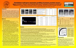
The principle of DNA microarrays • The differential gene expression assay between 2 tissue samples using the DNA microarray technology : normal or reference RNA vs cancer or test RNA • Total cellular mRNA is reversed transcribed to cDNA in the presence of 2 differentially fluor-labelled nucleotides (UTP) : cDNA molecules with either a green or a red fluoresence tag
These tagged cDNAs are then combined and hybridized to the microarray plate : containing tens of thousands of immobilized short DNA fragments of specified sequences • After hybridization and washing to remove nonspecific binding, the DNA microarray plate is subjected to excitation with lasers of different wavelengths : leading to distinct emissions from the Cy3 and Cy5 labelled probes
After detection by microscope scanning, the separate image files are exported to analysis software : they are converted to pseudo-colored images and a visual interpretation of expression changes is provided by merging the 2 images • The data are subjected to analysis with specific bioinformatic tools
These gene expression data • Referred to as signatures : because the expression patterns are distinct and unique for each type of tumor • Can be used to histologically classify similar tumors into specific subtypes • Providing clinically novel and relevant information
Reflecting the origin and function of different cell types • cf. Histochemical approaches : only discriminate between malignant and nonmalignant cells on the basis of morphologic appearance • Anticipated to provide in the immediate future a more accurate prognosis and prediction of response to individual therapy
The prognosis of cervical & endometrial cancer • The major conventional parameters • Clinical and/or surgical stage • Size and grade of tumor • Histologic type • Lymphatic spread • Vascular invasion
Additional parameters • Cytogenetic abnormalities • Acquired mutations of several proto-oncogenes, tumor suppressor genes, other cell cycle regulatory molecules -> Considered as candidate molecular events in the pathogenesis and the eventual evolution of the tumor to metastasis
Human papilloma virus (HPV) • The role of HPV infection : the development of preinvasive or invasive carcinoma of the cervix • Recent studies that use microarrays • HPV 16 E6 oncoprotein : Regulate differentiation-associated genes
HPV 16 E7 protein : regulate several key modulators • Signaling factors • Cell cycle regulators • Chaperones : Escape immune surveillance • E2 viral protein : Delay mitotic progression in HPV-mediated tumorigenesis
The recently established microarray DNA technology • The detection and typing of HPV infection • Sensitive high-throughput screening test for the detection of latent HPV • Essential insights on the mechanisms of multiple infections and the various genotypes of HPV
Single gene-based approaches for the investigation of endometrial carcinomas
No common molecular parameters have been formulated so far in endometrial cancer • K-ras proto-oncogene mutation • No prognostic value : Because, no correlation to • The stage • The histologic type • The grade of the tumor
Early endometrial cancer : Specific for detecting submicroscopic myometrial strips infiltrated with tumor cells • COX-2 expression • Lymph node metastasis • Parametrial invasion
Early studies : a very limited research • The identification of genes related to radiosensitiveity of cervical squamous cell carcinomas • The identification of genes involved in the development of cervical carcinomas • The identification of lovostatin-induced apoptosis-specific genes
The characterization of genes that are transactivated by the PTEN tumor suppressor gene in endometrial cells # Limited number of samples # Small number of genes contained in the cDNA microarrays → not provided any conclusive pattern of expression or gene signature for the neoplasia → not clarified the precise genes and the various steps of carcinogenesis
New studies : used cDNA microarrays : Systematic analysis of the pattern of expression during the various steps of cervical tumorigensis • Specific patterns of gene expression assigned to several cellular processes ① establish early enough during carcinogenesis ② can discriminate normal cervical tissue and LSIL from HSIL and cervical cancers
Many of these genes are also expressed in stroma adjacent to the cancer tissue • The extent of gene overexpression is increased during the progression from LSIL to HSIL and finally to cancer → The identification of reliable biomarkers associated with every stage of tumor progression → Eventually, leading to the improvement of early detection
4 major histologic types of ovarian carcinoma • Clear cell • Endometrioid • Mucinous • Serous → Directly analyzed using oligonucleotide microarrays
Expression profiling patterns • Mucinous and clear cell types can be readily distinguished from serous type, regardless of tumor stage and type • Clear cell carcinomas seem to be more similar to a subset of mucinous and endometrioid carcinomas than to serous carcinomas • Endometrioid carcinmas exhibit extensive overlap with the other histologic types
Clear cell ovarian carcinomas exhibit an expression profile distinct from other poor prognosis types : the 73 genes upregulated in clear cell → consistent with the capacity of being chemotherapy-resistant → hence of poor prognosis
Feasible pattern distinction between normal and ovarian cancer
Alternative ovarian cancer model • The clear distinction between normal human ovarian surface epithelial (HOSE) cell and epithelial ovarian carcinoma (EOC) • Based on primary cells expanded in vitro either from normal ovaries or from EOC • The elucidation of the molecular events occurring at specific cell types and stages of ovarian carcinogenesis
The set of differentially expressed genes in the epithelial ovarian carcinoma cell from this study : previously associated with ovarian tumorigenesis : novel genes -> in embryonic pattern formation • Mxil (a single tumor suppressor gene) : tissue-specific expression and upregulation in normal ovarian cell : downregulation in the EOC cell
The continuous passage and the nonreproducible culture conditions : tumor cell lines may not reflect the actual biologic events of the primary tumor • The loss of tumor markers during culture • The possibility of propagating selected subpopulation from the original primary tissue • The putative modification of gene expression by the in vitro expansion conditions
The choice of normal ovarian control : A relevant critical issue in correctly identifying the differentially expressed genes in tumors • The selection of the type of the normal control cell type can influence the set of differentially expressed genes with a tumor sample • The lack of a universal control tissue sample each time does not permit meaningful comparisons among similar studies • The type of normal tissue should be clearly specified
Lack of available early malignant ovarian tissues : A specific problem for a systematic analysis of the biologic phenomena of the early ovarian carcinogenesis • Most of the ovarian tumors are discovered when they have already proceeded beyond stage I • The required tissue size for the extraction of 10 to 50ug of total RNA for gene expression analysis exceeds the available amount of premalignant ovarian tissue
To address these difficulties • The short-term in vitro expansion of normal and malignant ovarian epithelial cells before RNA harvesting • The purification of ovarian epithelium • The amplification of the RNA
Genome-wide examination of chromosome : The expression patterns of the different stages of tumor progression of ovarian cancer • The patterns of 21 early stage : 17 late stage -> compared by using cDNA microarray analysis, comparative genomic hybridization • Early stage : stage I/II, endometrial or serous carcinomas • Late stage : stage III/IV, serous carcinomas
A more recent study : Identification of gene expression patterns of the 2 survival group • Advanced stage (III or IV), serous ovarian cancer -> short (< 2yr) : long (> 7yr) survival • Significant number of differentially expressed genes : IL2 receptor, chemokine ligands (CCL4, CCL5), several interferon pathway activities : immune system functions : upregulation -> a favorable outcome
The pattern of aberrant expression in the early stages of ovarian carcinogenesis : The potential for metastasis is an inherent feature of early stage cancers • Important implication on future strategies for rational treatment and screening • Aggressive treatment of poorly differentiated stage I ovarian cancers • The rationale for screening strategies
These tumors seem • to include subpopulations of cells exhibiting tissue-specific profiles predicting the site of metastasis • to argue against the current model that metastasis is derived from rare cells residing within the tumor • These new data alter radically our current model of tumor progression and metastasis : Sequential accumulation of numerous mutations occurring on a rare subpopulation of tumor cells
The prediction of response to chemotherapy : Recent studies have addressed the feasibility of generating novel reliable molecular markers using gene expression profiling • The paclitaxel-induced apoptosis pathways • The p53-independent mitochondrial pathway • The stress reaction-induced pathway -> Suppression of these pathways can contribute to the acquisition of resistance to paclitaxel
Epothilone B • Nontaxane agent being active in paclitaxed-resistant cells • The pattern of expression of ovarian cancer cell to Epothilone B : triggering of stress-related signal transduction pathways associated to TNF-a • This finding provides the impetus for further studies to delineate the mechanisms of drug resistance
Microarray analysis -> to characterize more efficiently human tumors at the gene expression level • Revealing significantly and highly altered genes between tumor and normal tissue -> to begin deciphering the pathways mostly affected in disease process
The diversification of the microarray platform expanding beyond DNA such as • proteins • carbohydrates • peptides • nanotube precursors -> to add a significant dimension to our understanding of the complexity of cellular machinery