Download

1 / 36
1.05k likes | 2.34k Views
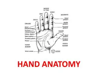
HAND ANATOMY. Dorsum Of Hand. Skin Cutaneous innervation Terminal branches of Radial Nerve Dorsal branch ulnar nerve Dorsal Venous network, arch proximal to MCH Drain from volar aspect as well Pressure of gripping does not impede the venous return Radial Side Cephalic Vein

E N D
Dorsum Of Hand • Skin • Cutaneous innervation Terminal branches of Radial Nerve Dorsal branch ulnar nerve • Dorsal Venous network, arch proximal to MCH Drain from volar aspect as well Pressure of gripping does not impede the venous return Radial Side Cephalic Vein Ulnar Side Basilic Vein
Palm of the Hand • Skin – Flexion creases and papillary ridges • Possibly improve the grip and increase the surface area • Sweat glands abound, No sebaceous glands • Ulnar Nv. Little ,½ Ring and Hypothenar eminence • Median Nv. Thumb, Index Middle, ½ Ring and Thenar eminence
Palm Fascia • Palmar Aponeurosis • Flexor Retinaculum, transverse carpal ligament Radial - Scaphoid tubercle, ridge of Trapezium Ulnar – Pisiform, hook of Hamate • Superficial relations, ulnar to radial Ulnar Nv., Ulnar Art., Covered by fascia giving origin to the hypothenar ms. – Guyon’s canal Palmar br. Ulnar nv , PL tendon, Palmar br. Median nv, Superficial palmar br. Radial art.
Palmar Spaces Thenar and Mid Palmar Spaces – Located dorsal to FT and volar to MC and Int. ms. Fascia Midpalmar oblique Septum Thenar Space between the thenar eminence and third metacarpal. Extends dorsally IbI the Int. ms. And Add. Pollicis .Mostly contains the first lumbrical Midpalmar Space IbI thenar and hypothenar space overlying the 3,4,5 MC Hypothenar Space Dorsal Sub aponeurotic Space Interdigital Web Space Radial,Ulnar bursa,Parona’s
Blood Supply • Radial Artery • Ulnar Artery • Superficial Palmar Arch • Deep palmar Arch • Anterior carpal arch • Posterior carpal arch
Radial Artery • Wrist - emerges medial to the brachioradialis • Superficial palmar branch – Superficial palmar arch • Dorsally IbI the radial carpal ligament and the APL EPB to emerge in the snuff box • Branches - Radial digital collateral artery Dorsal radial carpal branch, FDMA • Reenters palm IbI the two heads of 1st DI • A. radialis indices and A. princeps pollicis • Emerges IbI the transverse and oblique heads of adductor pollicis – Deep palmar arch • Post. Carpal Arch – DRCB, Ulnar A., Int. A. – 2, 3, 4 DMAs
Ulnar Artery and Nerve • Deep and radial to Ulnar nv and FCU • Superficial br.- Superficial palmar arch • Deep br.- Deep palmar arch • Ulnar nv ulnar and more superficial • Superficial br. Ulnar side of little and common digital nv to the little and ring • Deep br. Supplies the hypothenar muscles Curves around the hook of hamate and pierces the opponens digiti minimi along with the deep br. Ulnar A. to supply the 3,4th lumbricals and all interossei to end in the Adductor pollicis
Median Nerve • Enters the palm through the carpal tunnel • Three branches • Medial – Common digital to the ring and middle and common digital to the middle and index – gives a br to the second lumbrical • Lateral – Radial digital to the index and the whole of thumb – gives a br to the first lumbrical • Recurrent br./ muscular br. – thenar muscles
Thenar Eminence • Abductor Pollicis Brevis – arises FR and scaphoid tubercle inserts radial side base of proximal phalanx • Flexor Pollicis Brevis – arises FR and trapezium inserts radial sesmoid and radial border proximal phalanx – deep head ulnar nv • Opponens Pollicis – arises FR and trapezium inserts radial border metacarpal • Adductor Pollicis – arises 3rd MC transverse head, capitate oblique head inserts ulnar sesmoid and ulnar side base of proximal phalanx – Ulnar nv
Hypothenar Eminence • Abductor Digiti Minimi – arises FR and pisiform inserts ulnar side proximal phalanx and ext. expansion • Flexor Digiti Minimi Brevis – arises FR inserts base of proximal phalanx • Opponens Digiti Minimi – arises FR and hook of hamate inserts ulnar border 5th metacarpal
Flexor Tendons Flexor Digitorum Profundus Flexor Digitorum Superficialis Chiasma
Extensor Retinaculum • Ribbon like band <2.5 cm wide • Oblique across dorsal surface wrist joint • Medial attach.Radius anterolateral border • Lateral attach. Pisiform and Triquetral passes below the styloid process ulna • If attached to both the forearm bones the ER would be 30 % longer in pronation • Being oblique it is able to maintain a constant tension throughout the motion
Divide into six compartments by fibrous septa to the bone • Separate synovial sheaths for all the tendons except the EDC and EI
Nail Anatomy Perionychium – Nail Bed, Nail Fold, Eponychium, Paronychium, Hyponychium Nail Bed – Germinal matrix, sterile matrix Nail Fold – Dorsal roof, Ventral floor – germinal matrix – Lunula Germinal matrix produces 90% of the nail, sterile matrix adds inner layer which keeps the nail adherent, dorsal roof gives the shine