Download

1 / 36
360 likes | 400 Views
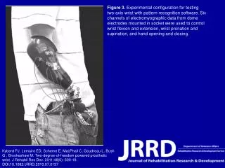
Hand & Wrist Skeletal Anatomy. Sports Medicine. Bellwork. Complete only the B portion of the BLT chart writing what you know about the hand and wrist. Then put BLT chart aside until the end of the lesson.

E N D
Hand & Wrist Skeletal Anatomy Sports Medicine Property of CTE Joint Venture
Bellwork • Complete only the B portion of the BLT chart writing what you know about the hand and wrist. Then put BLT chart aside until the end of the lesson. • Write-Pair-Share a list of all the anatomical structures of the hand that you can recall from previous lessons. Record list on paper and share with partner. Property of CTE Joint Venture
Objectives • Recognize skeletal anatomy for the hand & wrist • Recognize ligaments of the hand & wrist Property of CTE Joint Venture
Terminology • Carpals: small bones of the hand • Metacarpals: long bones of the hand, between the carpal bones and the phalanges • Phalanges: bones of the fingers Property of CTE Joint Venture
Bones of the Hand & Wrist • Ulna • Radius • Carpals (8) • Metacarpals (5) • Phalanges (14) • Distal • Intermediate • Proximal http://www.sports-injury-info.com/hand-anatomy.html Property of CTE Joint Venture
Bones of the Wrist • Ulna • Styloid Process • Radius • Styloid Process https://www.google.com/url?sa=i&rct=j&q=&esrc=s&source=images&cd=&cad=rja&uact=8&ved=0ahUKEwjJs8nWmKzUAhVp0FQKHY2nBKMQjhwIBQ&url=https%3A%2F%2Fplasticsurgerykey.com%2Fmanagement-of-wrist-fractures%2F&psig=AFQjCNE55xqAwlryP3FxyKP_UJQsgKC7BQ&ust=1496940591745748 Property of CTE Joint Venture
Carpal Bones • (Starting with distal row, lateral to medial) • 1. Trapezium • 2. Trapezoid • 3. Capitate • 4. Hamate • 5. Pisiform • 6. Scaphoid • 7. Lunate • 8. Triquetrum 2 3 4 1 5 6 8 7 Property of CTE Joint Venture
Metacarpals • Numbered from 1 to 5, starting with the thumb • 1 – thumb • 2 – pointer finger • 3 – middle finger • 4 – ring finger • 5 – pinkie finger 3 2 4 5 1 Property of CTE Joint Venture
Phalanges (14) • Proximal phalynx • Intermediate phalynx (except thumb) • Distal Phalynx Property of CTE Joint Venture
Identify the bones of the hand Property of CTE Joint Venture http://www.yalemedicalgroup.org/stw/Page.asp?PageID=STW023547
Ligaments of the Wrist • Wrist: • Ulnar collateral ligament • Radial collateral ligament • Transverse carpal ligament Property of CTE Joint Venture
Ligaments of the Hand • Fingers: • Collateral ligaments Property of CTE Joint Venture
Articulations of the wrist • Radiocarpal joints • Intercarpal joints • Carpometacarpal joints
Joints of the Hand & Wrist • Radiocarpal joint (wrist joint) • Carpal joints • Metacarpophalangeal joints • Phalangeal joints • Proximal interphalangeal (PIP) joint • Distal interphalangeal (DIP) joint
Radiocarpal Joint • The articulation between the radius and the carpal bones • Type of joint: condyloid • Allows flexion, extension, ulnar and radial deviation, and circumduction
Carpal Joints • Type of joint: gliding • Stabilized by various ligaments • Allow limited movement
Metacarpophalangeal Joints • Articulation between the metacarpals and the phalanges • 1st MCP (thumb) is a saddle joint • Allows flexion, extension, abduction, adduction and rotation
Metacarpophalangeal Joints • 2nd through 5th MCP joints are condyloid joints • Allow flexion, extension, abduction, adduction and circumduction
Interphalangeal Joints • Type of joint: hinge • Allow flexion and extension • Two categories of phalangeal joints: • proximal • distal
Interphalangeal Joints • Proximal interphalangeal (PIP) joint is the articulation between the proximal & intermediate phalanges • Distal interphalangeal (DIP) joint is the articulation between the intermediate and distal phalanges • Exception: The thumb only has an interphalangeal joint (IP) joint because it has just 2 phalanges
Carpal Ligaments • Wrist is stabilized by anterior, posterior, and interosseous ligaments. • Play important role in stabilization of the wrist complex. • Identification of specific ligament difficult if not impossible.
Bone structure allows 6 different motions Flexion Extension Adduction (Ulnar deviation) Abduction (Radial deviation) Pronation Supination Wrist Motion
Muscles of the Wrist • Wrist flexors: • group of muscles located on the anterior part of the wrist • perform flexion of the wrist
Wrist Flexors • Pronator Teres: strongest wrist & forearm pronator; works with the pronator quadratus • Flexor carpi radialis: flexes & abducts the wrist toward the radial side (radial deviation) • Flexor carpi ulnaris: flexes & adducts the wrist toward the ulnar side (ulnar deviation) • Palmaris longus: a secondary muscle active during wrist flexion • 14% OF PEOPLE DON’T HAVE ONE! DO YOU? • Oppose your pinkie & thumb, then slightly flex your wrist. Are two tendons popping up or just one? If just one, then you’re unique! • Supinator: performs wrist & forearm supination *Carpus is Latin for wrist!
Wrist Extensors • Wrist extensors: • group of muscles located on the posterior part of the wrist • perform extension of the wrist • Extensor carpi ulnaris: extends & adducts the wrist • Extensor carpi radialis longus & brevis: extend & abduct the wrist
Muscles of the Fingers • Finger flexors: • muscles on the anterior part of the hand • perform flexion of the fingers • Finger extensors: • muscles on the posterior part of the hand • perform extension of the fingers
Finger Flexors • Flexor digitorum superficialis: Flexes the digits at the proximal interphalangeal joints • Flexor digitorum profundus: Flexes the digits at the distal interphalangeal joints • Flexor pollicis longus: Flexes the thumb at the IP joint • Flexor pollicis brevis: Flexes the thumb at the MCP joint
Finger Extensors • Extensor digitiminimi • Extends the pinkie finger • Extensor indicis • Extends the index finger • Extensor digitorum • Extends the fingers as a group • Extensor pollicis brevis • Extends the thumb at the MCP joint • Extensor pollicis longus • Extends the thumb at the IP joint • Abductor pollicis • Abducts the thumb
Flexor Retinaculum • Strong fibrous band that restrains or holds in place tissue, muscles or organs • Restraining structure for the flexor tendons
Extensor retinaculum • Strong fibrous band • Restraining structure for the extensor tendons
Review Property of CTE Joint Venture
NumberedHeads 4 Students Number 1-4 Identify the skeletal structure that the teacher is pointing to on the model of the hand and wrist. Property of CTE Joint Venture PROPERTY OF PIMA COUNTY JTED, 2010
Closure • Write down the anatomical names on a piece of paper that your teacher points to • Share your answers with a partner until you are sure you agree on the anatomy • Finish your “L” and “T” columns on your BLT sheet Property of CTE Joint Venture