Download
1 / 36
360 likes | 498 Views
Beamline 8.3.1 summary. Strong PRT and staff Robust optics and endstation Safety: stable, simple operations Funding: Operational funding secure Scientific productivity: high Future: streamlining success. ALS beamline 8.3.1. Diffraction Methods Research.

E N D
Beamline 8.3.1 summary Strong PRT and staff Robust optics and endstation Safety: stable, simple operations Funding: Operational funding secure Scientific productivity: high Future: streamlining success
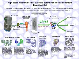
ALS beamline 8.3.1 Diffraction Methods Research Holton J. M. (2009) J. Synchrotron Rad.16 133-42
Howells et al. (2009) J. Electron. Spectrosc. Relat. Phenom.170 4-12 103 100 maximum tolerable dose (MGy) 10 1 1 2 3 5 7 10 20 40 70 100 resolution (Å)
10 MGy/Å what the is a MGy? http://bl831.als.lbl.gov/ damage_rates.pdf Holton J. M. (2009) J. Synchrotron Rad.16 133-42
Radiation Damage Model normalized total intensity accumulated dose (MGy)
Radiation Damage Model slopes (Å2/MGy): lysozyme: 0.013 apoferritin: 0.016 best-fit B factor Kmetko et. al. (2006): lysozyme: 0.012 apoferritin: 0.017 accumulated dose (MGy)
simulated real Simulated diffraction imageMLFSOM
Glaeser et.al. (2000) Sliz et.al. (2003) 1 μm amyloids Nelson et al. 2005 Sawaya et al. 2007 ~12 μm xylanase Moukhametzianov et al. 2008 5 μm cypovirus polyhedra Coulibaly et. al. 2007 predicted Glaeser et.al. (2000) 5 μm (13x) bovine rhodopsin Standfuss et al. 2007 theoretical Crystal Size http://bl831.als.lbl.gov/~jamesh/xtalsize.html CC to correct model crystal size (μm)
Minimum Crystal Size http://bl831.als.lbl.gov/~jamesh/xtalsize.html MW VM2 nxtal = n0 ℓxℓyℓz (d3-1.53) exp(-0.5 B/d2) nxtal - number of crystals needed n0 - empirical constant (~ 3) MW - molecular weight (kDa) VM - Matthews number (~2.5 Å3/Da) ℓ - crystal size (microns) d - d-spacing of interest (Å) B - Wilson B factor (Å2) B ≈ 4 d2 + 12 Holton J. M. (2009) J. Synchrotron Rad.16 133-42
Theoretical limit: Where: IDL - average damage-limited intensity (photons/hkl) at a given resolution 105 - converting R from μm to m, re from m to Å, ρ from g/cm3 to kg/m3 and MGy to Gy re - classical electron radius (2.818 x 10⁻¹⁵ m/electron) h - Planck’s constant (6.626 x 10⁻³⁴ J∙s) c - speed of light (299792458 m/s) fdecayed - fractional progress toward completely faded spots at end of data set ρ - density of crystal (~1.2 g/cm3) R - radius of the spherical crystal (μm) λ - X-ray wavelength (Å) fNH - the Nave & Hill (2005) dose capture fraction (1 for large crystals) nASU - number of proteins in the asymmetric unit Mr - molecular weight of the protein (Daltons or g/mol) VM - Matthews’s coefficient (~2.4 Å3/Dalton) H - Howells’s criterion (10 MGy/Å) θ - Bragg angle a2 - number-averaged squared structure factor per protein atom (electron2) Ma - number-averaged atomic weight of a protein atom (~7.1 Daltons) B - average (Wilson) temperature factor (Å2) μ - attenuation coefficient of sphere material (m-1) μen - mass energy-absorption coefficient of sphere material (m-1) Holton J. M. and Frankel K. A. (2010) Acta Dsubmitted
Theoretical limit: for lysozyme photon spot μm3 1.0 at ~2.4 Å Holton J. M. and Frankel K. A. (2010) Acta Dsubmitted
Optimum exposure time(faint spots) thr optimum exposure time for data set (s) tref exposure time of reference image (s) bgref background level near weak spots on reference image (ADU) bg0 ADC offset of detector (ADU) σ0rms read-out noise (ADU) gain ADU/photon m multiplicity of data set (including partials) Short answer: bghr = 90 ADU for ADSC Q315r
Damage changes absorption spectrum 1 0 counts Photon energy (eV) Holton J. M. (2007) J. Synchrotron Rad.14 51-72
fluorescence probe for damage 25mM SeMet in 25% glycerol Exposing at 12680 eV Se cross-section at 12680 eV 0.0 0.2 0.4 0.6 0.8 1.0 Fraction unconverted 0 50 100 150 200 250 300 350 400 fluence (1015 photons/mm2) Holton J. M. (2007) J. Synchrotron Rad.14 51-72
fluorescence probe for damage Wide range of decay rates seen Half-dose = 5.5 ± 0.6 MGy 8 mM SeMet in NaOH Half-dose = 41.7 ± 4 MGy “GCN4” in crystal 0.0 0.2 0.4 0.6 0.8 1.0 Fraction unconverted Protection factor: 660% ± 94% 0 50 100 150 200 Absorbed Dose (MGy) Holton J. M. (2007) J. Synchrotron Rad.14 51-72
Take-home lesson: Best strategy: radiation damage to metal sites is unpredictable 5 MGy to complete data geometrically increasing exposure Holton J. M. (2007) J. Synchrotron Rad.14 51-72
Spatial Noise down up Rseparate
Spatial Noise odd even Rmixed
Spatial Noise separate: mixed: 2.5% 0.9% 2.5%2-0.9%2=2.3%2
Spatial Noise mult >(—)2 2.3% <ΔF/F>
Minimum Crystal Size http://bl831.als.lbl.gov/~jamesh/xtalsize.html MW VM2 nxtal = n0 ℓxℓyℓz (d3-1.53) exp(-0.5 B/d2) nxtal - number of crystals needed n0 - 3 for complete data set, 180 for MAD MW - molecular weight (kDa) VM - Matthews number (~2.5 Å3/Da) ℓ - crystal size (microns) d - d-spacing of interest (Å) B - Wilson B factor (Å2) B ≈ 4 d2 + 12 Holton J. M. (2009) J. Synchrotron Rad.16 133-42
Take-home lesson: Best strategy: need better crystals for MAD find them
accurate, unattended data colleciton
Re-centering beamline microscope reference image
accurate, unattended screening
Cu sample shadow on detector