Download

1 / 31
360 likes | 633 Views
Dr. Wajeeh Nama Hussein 2012. Acne mimickers Another cause for unresponsive acne. Sahar H. Al- Natour Department of Dermatology, College of Medicine, University of Dammam , Saudi Arabia King Fahad Hospital of the University, Al- Khobar , Saudi Arabia

E N D
Dr. Wajeeh Nama Hussein 2012 Acne mimickersAnother cause for unresponsive acne
Sahar H. Al-Natour • Department of Dermatology, College of Medicine, University of Dammam, Saudi Arabia • King Fahad Hospital of the University, Al-Khobar, Saudi Arabia • Journal of the Saudi Society of Dermatology & Dermatologic Surgery (2012)
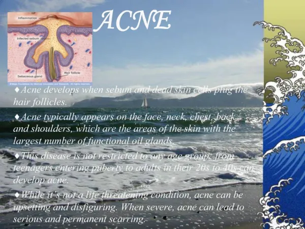
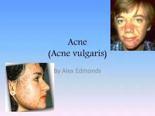
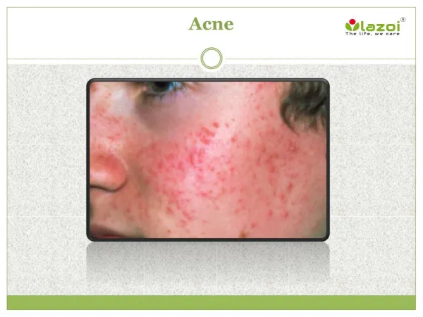
Introduction • Acne is a worldwide disorder affecting millions of patients each year. It is sufficiently common that it has been termed physiological. • Most of the presentations of acne are pleopmorphic, manifesting with a variety of lesions consisting of comedones, papules, pustules, nodules, and pitted and hypertrophic scars. Lack of response despite an effective treatment regimen and good compliance should alert the treating physician to consider all possible scenarios. One of the main reasons for unresponsiveness is the misdiagnosis due to the presence of conditions mimicking acne.
Conditions likely to be mistaken for acne • Conditions presenting with comedones • Nevus comedonicus • Favre Racouchot Syndrome • Conditions with acne in their name • Acne rosacea, var. perioral dermatitis • Acne agminata • Chloracne • Drug induced acne
Conditions likely to be mistaken for acne • Appendageal abnormalities • Sebaceous hyperplasia • Miliaria • Milia • Benign tumors • Syringoma • Trichoepithelioma • Angiofibroma (adenoma sebaceum) • Conditions in the neonate • Neonatal cephalic pustulos
Conditions likely to be mistaken for acne • Conditions with a follicular component • Folliculitis • Gram negative folliculitis • Eosinophilicfolliculitis • Follicular mycoses fungoides • Pseudofolliculitisbarbae • Keratosispilarisfaceii
1. Nevus comedonicus • A benign and rare hamartoma of the pilosebaceous unit present at birth before the age of 10 years. Clinically, this nevus presents as grouped comedo-like lesions in a single circumscribed area or in a linear array most commonly on the face, trunk, neck or upper extremities. Bilateral and segmental forms have also been described and non-hairy sites can be affected. • When present, other abnormalities should be looked for such as cataracts, skeletal abnormalities or developmental delays. Histologically, these nevi consist of underdeveloped hair shafts and epidermal invaginations containing a keratin plug
2. Favre Racouchat Syndrome (Nodular elasotosis with cysts and comedones) • Is a disorder consisting of multiple open and closed comedones in the background of actinically damaged skin. Characterized by the presence of multiple large persistent open and closed comedones in a background of solar elastosis mainly on the lateral and inferior aspects of the periorbital areas in middle aged or elderly patients . • Marked actinic damage presenting as yellowish leathery skin particularly of the cheeks, yellowish nodules of the periorbital areas and wrinkling is characteristic. No infl-ammation is present, unlike most comedones seen in acne vulgaris . Long term exposure to sunlight and heavy smoking are the main predisposing factors. Histologically, there are dilated pilosebaceous openings and cyst-like spaces filled with horny elastotic material.
3. Rosacea (Acne rosacea) • Although both conditions (acne and rosacea) present with papules and pustules, the papules of rosacea are asymptomatic, rose colored, and dome shaped rather than the painful pointed red papules of acne. In rosacea, the lesions occur typically in the mid line and convex surfaces of the faces. Comedones are characteristically absent. The vascular component of rosacea presents as transient flushing precipitated by hot drinks, spicy foods, heat, emotion, and other causes of rapid body temperature changes early in the disease later becoming a persistent erythema with telengiectatic vessels. Other characteristics of rosacea are edema, coarseness of the skin and phymatous changes. Extracutaneous associations include ocular manifestations.
4. Perioral dermatitis • Perioral dermatitis affects young women especially those who have been using moderate and strong fluorinated steroids on the face and presents as small clustered itchy or tender red papules in the perioral area which characteristically spare the vermillion border of the lips. The skin is typically erythematous, dry and scaly around the nose. Periorbital eruption is not unusual. Perioral and periorbital dermatitis often appear in patients with vascular rosacea. The link to rosacea is not certain but both share similar histopathologicalfindings. The main differentiating feature of perioral dermatitis from acne vulgaris is the absence of comedones and the free zone along the vermillion border of the lips in the former.
5. Acne agminata (lupus miliarisdissaminatusfaciei) • Lupus miliarisdissaminatusfaciei (LMDF) is a rare disease affecting the face primarily in young and middle aged adults. Skowron et al. proposed a name change from LMDF to FIGURE (facial idiopathic granulomas with regressive evolution) in 2000; to date this name change has not been widely accepted. • Clinically, asymptomatic discrete monomorphous yellow brown papules appear in the center of the face with a predilection to the periocular area. Comedones are typically absent. The diagnosis is established by histological examination. Well definedcaseation necrosis, multinucleated giant cells and epithelioid cells are seen in the dermis and are diagnostic. Spontaneous regression with mild scarring is the expected course.
6. Chloracne • Chloracne is a rare acne-like skin condition resulting from occupational and environmental exposure to halogenated polycyclic hydrocarbons. The eruption may not appear for three to four weeks after toxic exposure in some instances, but may appear within days in the event of massive exposure in others. Chloracne is distinguished from acne vulgaris by the predominance of large open comedones over closed comedones typically concentrated over the cheeks, malar crescent and retroauricular folds. Other body sites can also be affected mainly by the armpits, groins, scrotum, buttocks and thighs. Uninflammed nodules and cystic lesions are usually seen accompanying the open comedones.
7. Drug induced acne • An abrupt eruption consisting of monomorphous follicular papules and pustules is typically seen over the chest in adolescents or adults on moderate to high doses of oral corticosteroids for several weeks in contrast to the pleomorphic morphology of lesions seen in acne vulgaris. • The main differentiating features from acne are the absence of comedones and the associated itching. Other major drugs implicated in acneiform eruptions include glucocorticoids, androgens, anti-epileptic drugs, iodides, and isoniazids , and oral contraceptives containing progestins with androgenic properties. The recent introduction of (EGFR Inhibitors) has led dermatologists to see follicular eruptions with increasing frequency. Less commonly, azathioprine can be implicated as the underlying cause of acneiform eruptions.
8. Sebaceous hyperplasia • Sebaceous hyperplasia is common and presents as one or several yellowish papules usually over the forehead and cheeks but can also present over the upper trunk and genitalia. The clue to the diagnosis is the presence of a central hair follicle surrounded by yellowish lobules. Comedones are typically absent. Risk groups for the development of sebaceous hyperplasia are those with immunosuppression, transplant patients or in Muir Torre Syndrome . Diascopy is a useful non-invasive diagnostic tool that aids in the clinical diagnosis and in distinguishing between nodular basal cell carcinoma (BCC) and sebaceous hyperplasia demonstrating the prominent blood vessels in sebaceous hyperplasia . A skin biopsy is indicated in some cases mainly to exclude BCC
9. Miliaria • Miliariarubra, the most common clinical form of miliaria can be confused with acne. This eruption typically occurs after days to weeks of exposure to hot and humid environment as small nonfollicular, erythematousmacules and papules 1–3 mm in diameter topped by a punctuate vesicle over the neck and trunk of both children and adults. The eruption is typically associated with pruritus. Miliariarubra may become pustular in chronic and severe cases and when so, it is referred to as miliariarubrapustulosa. The contents of the pustules are usually sterile bacteriologically, but non-pathogenic cocci may be present. This eruption is differentiated from acne vulgaris by the absence of comedones and positive history of excessive sweating.
10. Milium • Milia are common small epidermoid cysts that can occur at any age. Milia present clinically as asymptomatic white or yellow subepidermal papules 1–2 mm in diameter. Between 40% and 50% of all infants are found to have milia on the face par-ticularly on the nose that resolves spontaneously within 4 weeks of life. They may also be found on the mucosa (Epstein pearls) and palate (Bohn nodules). Milia may occur as a primary phenomenon on the face or secondary to blistering processes, superficial ulceration from either cosmetic proce-dures or trauma or as a result of corticosteroid induced atrophy. Secondary milia are found anywhere on the body at the sites affected by the predisposing condition.
11. Syringoma • Syringoma is a benign adnexal neoplasm formed by well differentiated ductal elements. In syringomas, small asymptomatic translucent skin colored papules 1–3 mm in diameter, clustered but discrete appear almost exclusively on the eyelids and upper cheeks in adolescents and adult women mostly. However, in • rare cases they can be associated with pruritus in the setting of severe perspiration. Other sites of involvement which can easily be missed include the chest, lower abdomen and groins. Microscopically, the diagnostic pathology includes dilated cystic sweat ducts with the typical tad-pole shaped ducts in sclerotic stroma.
12. Trichoepithelioma • Trichoepithelioma is a benign adnexal neoplasm. Present as single or multiple asymptomatic papules or nodules usually less than 1 cm in diameter typically in the mid face occurring after puberty. The characteristic papule is firm, rounded shiny, slightly translucent ranging in color from yellow, pink, brown or blue. Most often the lesions are grouped but discrete. Comedones are absent. A positive family history is usually elicited. Skin biopsy confirms the diagnosis and differentiates trichoepithelioma from syringoma and BCC. The diagnostic pathology is the well circumscribed superficial lesion consisting of clusters of basaloid cells with a fibrousstroma. Immunohistochemistry helps in the differentiation of trichoepitheliomas from BCC.
13. Angiofibroma • Cutaneousangiofibroma refers to a group of lesions with different clinical presentations but with similar histology. Fibrous papules present as asymptomatic solitary skin colored to reddish dome shaped papules on the face of adults with a predilection on the nose. Multiple facial angiofibromas associated with tuberous sclerosis (TS) typically present as asymptomatic pink or skin-colored telengiectatic papules commonly observed over the cheeks, nose and nasolabial folds in a butterfly distribution.
These lesions typically appear between age 3 and 10 years and tend to increase in size and number in adolescence. Patients with TS can also have peri-ungalfibromas which appear later in life than the facial angiofibromas. Both of these lesions are considered as primary diagnostic features of TS. Other cutaneous signs pointing to the diagnosis include the ash leaf hypopigmentation and shagreen patch.
14. Folliculitis • Folliculitis is a very common disorder characterized by perifollicular pustules often arising on a base of erythema. Lesions are tender or painful but most commonly very pruritic. Folliculitis favors areas with terminal hair such as the scalp and beard, trunk, buttocks and thighs. Bacteriologically, these pus-tules are usually sterile, occasionally Staphylococcus aureus is isolated. When pustules are not present, perifollicularerythematous papules or a superimposed collarette of scales are a clue to the diagnosis.
15. Gram negative folliculitis • G-vefolliculitis presents as a rash consisting of tender pustular lesions with a few inflammatory papules and comedones over the facial T-zone and in a perinasal distribution mostly in adult men with oily skin. A typical history of long term use of tetracycline or topical antibiotics in patients with acne vulgaris is elicited in those with this eruption. G-vefolliculitis should also be considered in those acne patients who have a sudden flare-up of pustular lesions and in those whose acne fails to respond to acne treatment.
Bacterial cultures of the pustular contents demon-strate gram negative organisms: Klebsiella, Escherichia, Pro-eus, Serratia species. Cases with Citrobacter species have also been described. This condition can present as a real challenge in the diagnosis and management. Because this gram negative folliculitis occurs in patients with existing acne, the new eruption is often erroneously diagnosed as an exaccerbation of acne
16. Eosinophilicfolliculitis of HIV/AIDS • Itchy papules and pustules with some urticated lesions and ab-sence of comedones on the face, scalp and trunk with a dura-tion of weeks or months in the setting of HIV infection all point to a diagnosis of eosinophilicfolliculitis. The sparing of the palms and soles differentiates Eosinophilicfolliculitis (EF) from Ofuji’s disease. EF presents with elevated IgE levels, eosinophilia and peripheral leukocytosis. A CD4 count below 300 cells/mm3 confirms the diagnosis. Histologically, spongiosis and exocytosis of eosino-phils and lymphocytes into the follicular epithelium are demonstrated.
Eosinophilicpustularfolliculitis (Ofuji’s disease) • can also occur in bone marrow transplant patients and in inherited immunodeficiencies. The typical eruption is characterized by recurrent crops of intensely pruritic large follicular papules and pustules with peripheral extension and central clearing typically over the ‘‘acne prone’’ sites of the body and involving the palms and soles.
17. Folliculotropic mycoses fungoides • Patients present with grouped asymptomatic follicular papules, pustules, infiltratedcomedone-like lesions and infiltratederythematous plaques on the face and neck but also of the trunk. An associated alopecia is quite common. Infiltrated plaques over the eyebrow with concurrent alopecia is also a common presentation of this variant of mycoses fungoides. True comedones are absent. Histologically, folliculotropism rather than epidermotropism of lymphocytes and atypical hyperchromatic T cells are evident in hematoxylin and eosin staining. Mucin deposits are demonstrated by Alcian blue staining. The distinctive clinical and histological features establish the diagnosis.
18. Pseudofolliculitisbarbae • Pseudofolliculitisbarbae is a common and painful eruption that occurs in the beard area of men who shave. This condition favors individuals with curly hairs and those of African descent. This acneiform eruption consists of perifollicularinflammatory papules and pustules with ingrown hairs in the beard and neck areas. If the hair shaft is lifted up, the free end of the hair is seen to emerge from the papule. Considerable cosmetic disfigurement may occur with the chronicity of the folliculits causing post-inflammatory pigmentation, scarring and keloid formation. Comedones are absent.
19. Keratosispilarisatrophicansfaceii(Ulerythemaophryogenes) • Keratosispilarisatrophicansfaceii is an autosomal dominant (AD) condition presenting in infancy with erythematous follicular papules with central keratotic plugs which eventually develop follicular atrophy. The typical cutaneous presentation is a scarring alopecia of the outer third of the eyebrow mixed with tiny rough follicular lesions. Other common sites are the lips, cheeks and forhead. The rough follicular spots on a base of erythema over the cheeks can be mistaken for comedones.
20. Neonatal cephalic pustulosis • A transient eruption that occurs in 20% of healthy newborns. Typically, small inflamed papules and pustules without comedones appear on the cheeks and nasal bridge. Several species of Malassezia have been proposed as the etiologic factors of this eruption. The response of this eruption to 2% ketoconazole cream gives additional support to this theory. Spontaneous involution over a few weeks to months is expected.
Conclusion • When confronted with a non responding acneiform eruption despite an adequate treatment regimen in a compliant patient, the possibility of acne mimickers should be considered.