Download
1 / 49
540 likes | 1.1k Views
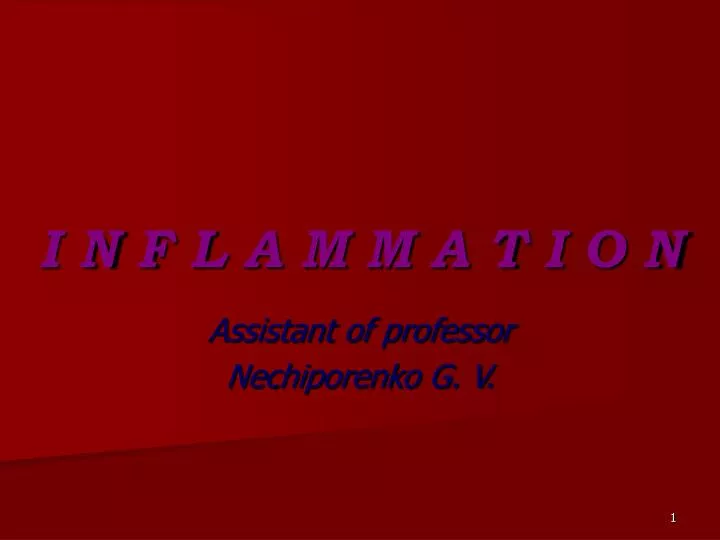
I N F L A M M A T I O N. Assistant of professor Nechiporenko G. V. Inflammation - is a local vascular-mesenchymal reaction to injury of living tissue due to different agents. This reaction has protective-adaptive character. It can kill agent caused injury and repair injured tissue.

E N D
I N F L A M M A T I O N Assistant of professor Nechiporenko G. V.
Inflammation- is a local vascular-mesenchymal reaction to injury of living tissue due to different agents. • This reaction has protective-adaptive character. • It can kill agent caused injury and repair injured tissue. • Inflammation contains elements of pathology and physiology. • Heat (calor), redness (rubor), edema (tumor), pain (dolor), and loss of function (functio laesa).
Types of inflammation • Morphological types: alterative, exudative, and proliferative (productive) • Due to etiology: specific and non-specific (banal) • Due to duration: acute, subacute, and chronic • Due to type of tissue’s reaction: normergic, hyperergic and hypoergic
Cells marginated along the dilated venule wall (arrow) are squeezing through the basement membrane (diapedesis) and spilling out into extravascular space.
Exudative inflammation • Catarrhal • Serous • Purulent (suppurative) • Fibrinous • Hemorrhagic • Putrefactive • Mixed
Serous inflammation • It is characterized by exudate contained about 2% proteins, single neutrophilic polymorphs and mesothelial cells. It occurs in serosa, mucosa, meninges, skin and internal organs. • Outcome is usually favorable. In severe cases it can be progressive into serous-purulent.
Suppurative inflammation • Phlegmon- diffuse unbounded purulent inflammation. Types: soft and dense. • Abscess- local purulent inflammation with necrosis and formation of cavity. • Empyema- appearance of pus in serosal cavities or organs with cavity. • Furuncle- purulent inflammation of hair follicle. • Carbuncle- formation of several closely disposed furuncles with necrotic centre.
Pustule- purulent rash in epidermis. Pimple. • Panaritium- purulent inflammation of ungula phalanx. Panaris, whitlow, felon. • Paronychia- purulent inflammation of soft periungual tissue. • Pyemia- septic dissemination in blood. • Fistula- pathologically formed canal from organ to cavity, other organ or skin for pus excretion.
Here is abscess in the lung. The alveoli in that area are destroyed.
Acute Streptococcus meningitis.A purulent exudate is seen in the meninges. The exudate obscures the sulci.
Hand, healing by secondary intention A whitish-greenish-yellow neutrophilic exudate represents an inflammatory response to bacterial invasion of the wound.
Outcomes of purulent inflammation • Resolution, • scarring, • petrification, • organization, • incapsulation, • acute toxemia, • pyemia, • sepsis • keloid • chronic duration, • amyloidosis
Fibrinous inflammation • Exudate contains fibrin, neutrophils and necrotic tissue elements. • It occurs in mucosa, serosa, meninges, and internal organs (lungs). • Croupous and dyphtheritic types of fibrinous inflammation.
This yellow-green exudate on the surface of an inflamed, hyperemic bowel mucosa consists of many neutrophils along with fibrin and amorphous debris from dying cells.
Microscopically, exudate consists of inflammatory cells, necrotic epithelium, and mucus in which the overgrowth of microorganisms takes place. The underlying mucosa shows congested vessels, but is still intact.
Here, the pericardial cavity has been opened to reveal fibrinous pericarditis with strands of stringy pale fibrin between visceral and parietal pericardium.
The fibrinous exudate consists of pink strands of fibrin jutting from the pericardial surface at the upper left. Below this, there are a few scattered inflammatory cells.
Outcomes of fibrinous inflammation • Resolution • Organization • Ulcers • Scarring • Obliteration of cavity • Chronic duration
Miliary tuberculosis of the lung.The granulomas1-2 mm are scattered like millet seeds.
Here are two pulmonary granulomas in tuberculosis. They typically consist of epithelioid macrophages, giant cells, lymphocytes, plasma cells, and fibroblasts.
These are epithelioid cells around the center of a granuloma. They get their name from the fact that they have lots of pink cytoplasm similar to squamous epithelial cells. Their nuclei tend to be long and stringy.
Lepromatous leprosy. Large collections of foamy macrophages (Virchow cells) infiltrate the dermis.
Necrotizing granulomatous vasculitis (Wegener's granulomatosis).This is a renal biopsy with a focal lesion centered around a blood vessel.
Sarcoidosis,granulomatous inflammationinSpleen Granulomas are scattered diffusely throughout the splenic parenchyma.
Sarcoidosis, Lymph node The granulomas are closely packed, often merging, with scant intervening lymphoid tissue. Scattered multinucleated giant cells are apparent.
The granulomatous inflammation in Crohn's disease is demonstrated with epithelioid cells, giant cells, and many lymphocytes.
Disseminated histoplasmosis in liver. Many fungal infections can produce a granulomatous pattern. The immune response is often poor, so granulomas are poorly formed. This portion of liver demonstrates some pinpoint yellow-tan granulomas.
This is infection with Histoplasma capsulatum. Note how each macrophage is filled with numerous small organisms. The organisms have a clear zone around a central blue nucleus which gives the cell membrane the appearance of a capsule.
Sometimes the inflammatory reaction is mainly one of scarring, as seen here with a silicotic nodule of the lung. The inhaled silica persists indefinitely and produces an inflammatory reaction that is marked by prominent fibrosis. Dense pink collagen is seen in the center of the nodule.
The interstitial lymphocytic infiltrates with little necrosis are characteristic for a viral myocarditis (Coxsackie B). It may be a cause for sudden death in young persons.
Chronic inflammation of the bronchi has led to dilation and scarring with increased tan to white collagenous tissue.
The end result of inflammation can be scarring. Here, the alveolar walls are thickened and filled with pink collagen.
Liver, chronic inflammation, cirrhosis Secondary to a prior episode of severe hepatitis, fibrosis has developed in this liver, altering its architecture.