Download
1 / 59
590 likes | 605 Views
Chapter 2 Epithelium. What is epithelium?. Conception: Epithelium is a tissue composed of closely aggregated polyhedral cells and very little extracellular matrix. Distribution : Cells cover body surfaces, line body cavities, and constitute glands. 1.General feature:

E N D
What is epithelium? Conception: Epithelium is a tissue composed of closely aggregated polyhedral cells and very little extracellular matrix. Distribution: Cells cover body surfaces, line body cavities, and constitute glands.
1.General feature: 1) contain more cells and less extracellular ground substance 2) Polarisation(polarity): ---free outer surface: face air or other things ---basal surface: have basement membrane, to face underlying CT, --- lateral surface: between adjacent cells 3) Avascularity, but innervation: ---no blood vessels ---rich in nerve terminals 4) Having functions of protection, secretion, absorption and sensory reception
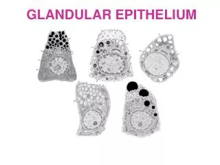
2.Classification of Epithelium 1)Covering epithelium: the epithelium which cover body surface or line the inner surface of body cavities, tubes and sac. 2)Glandular epithelium: the epithelium which main function is secretion. 3)Sensory epithelium: the epithelium which has special sensory function.
3. Classification of covering epithelium According to the number of layer and shape of cells Simple epi.: ---simple squamous epi. ---simple cuboidal epi. ---simple columnar epi. ---pseudostratified ciliated columnar epi. Stratified epi.: ---stratified squamous epi. ---stratified columnar epi. ---transitional epi.
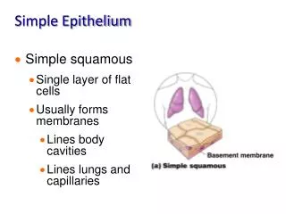
1)simple squamous epi: ---structural feature: /one layer flattened cells, cell border are interdigitate /with flattened ellipsoid nucleus
---distribution: Three types: 1.mesothelium:the simple squamous epi. which line the inner surface of body cavities such as thoracic, pericardiac and abdominal cavities. 2. endothelium:the simple squamous epi. which line the inner surface of cardiovascular and lymphatic system. 3. other place: alveoli, parietal layers of renal capsule.
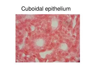
2) Simple cuboidal epi.: ---structural feature: • one layer of cells, with same height and width , hexagonal outline in surface view. • spherical centrally-located nucleus
---distribution: /the renal tubule /thyroid /the some ducts of glands ---function: covering and secretion renal tubule thyroid
3) Simple columnar epi.: ---structural features: • one layer of columnar cells, with basally located ovoid nucleus
---distribution: gastrointestinal tract gallbladder uterus ---function: secretion and absorption goblet cell: scattered, secreting granules-mucinogen granules-mucus goblet cell simple columnar epi
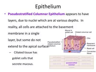
five types of cells 4) Pseudostratified ciliated columnar epi.: ---Structural feature: 1, Five types of cells columnar cell (ciliated); goblet cell fusiform cell; basal cell: pyramid-shaped diffuse neuroendocrine cell 2, Every cell locate on basement membrance: Simple epi.
---distribution: inner surface of large duct of respiratory trachea bronchi nasal The epithelium of trachea
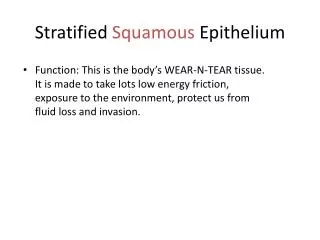
5) Stratified squamous epi.: Three layers ---structural features: 1. Deepest (basal) cells: one layer of cuboidal cells 2. The cells in intermediate regions: several layers of polygonal –shaped cells 3. To the surface: more and more flattened cells
---distributon: • non-keratinised: mouth, pharynx, esophagus, and vagina • keratinised: the surface of body, make up the skin non-keratinised keratinised
keratinised non-keratinised
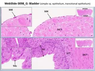
6) Transitional epi.: • flexible-including the number of layers and shape of cells • Two condition: • in the distended bladder: there are two to three layers of cells. The cells become flattened. • in the contracted bladder : there are six to seven layers of cells.
in the contracted bladder in the distendedbladder
---distribution:Urinary tract (ureters, bladder, urethra) The surface cells are very large and cuboidal in shape, covering several deep cells. Superficial cells
Summary 1 General feature: 1) contain more cells and less extracellular ground substance 2) Polarisation: ---free outer surface: face air or other things ---basal surface: have basement membrane, to face underlying CT, 3) ---no blood vessels ---rich in nerve terminals 4) Having many functions
Classification of covering epithelium According to the number of layer and shape of cells Simple epi.: ---simple squamous epi. ---simple cuboidal epi. ---simple columnar epi. ---pseudostratified ciliated columnar epi. Stratified epi.: ---stratified squamous epi. ---stratified columnar epi. ---transitional epi.
Exercise 1 for Histology • 1.The 4 basic types of tissue are ______________,______________,_________________and ________________. • 2. Describe the Classification of each covering epithelial type.
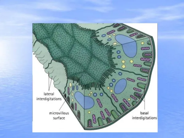
Specialisations of free surface specializations of the lateral surface specialization of basal surface
1)Specialisations of free surface Microvilli Cell coat Cilia
①microvilli: • defination:Microvilli are finger-like cytoplasmic projection on the free surface of most epithelial cells. • Distribution: striated border: intestinal epi. cell • brush border: proximal renal tubule
structure: 0.1um in diameter, with different longth. surface: cell membrane with cell coat core: longitudinal microfilament-actin filament fixed on terminal web terminal web: made up of transverse-arranged filament at the apical side of cells Function: increase the surface areas for absorption
②cell coat: ---defination: a thick layer of extracellular glycoprotein ---function: adherence, supporting, protection, exchange of material and recognize
③ cilia: ---defination: elongated, motile projections of cell membrane and cytoplasm protruding from free surface
---structure: • 5-10um long, 0.2um in diameter • surface: cell membrane • core: microtubules, 9X2+2 • basal body: centrioles-connected with microtubules
---function: swing to produce a forward-moving wave ---distribution: epithelial cells of respiratory tract respiratory tract
---intercellular connection of adjacent cells: • non-special: the minute space and cadherin(cell adherent molecules) • special: junctional structures
junctional structures Tight junction (zonula occludens) Intermediate junction (zonula adherens) Gap junction (communication junction) Desmosome (macula adherens)
①Tight junction (zonula occludens): ---structure: • apical part • point-liked fused between adjacent cells • form anastomosing network ---function: seal the space between cells
② intermediate junction (zonula adherens): ---structure: • below the tight junction • a gap of 15-20nm in width with medium electron-density filament material • plaque of electron-dense materials, with attached microfilament-make up of terminal web ---function: /adherens /keep the cell shape /transfer cell contract force
③gap junction (communicating junction): ---structure: • the smallest gap of 2-3 nm • connexons: -consist of protein -7~9nm in diameter -composed of 6-subunits of proteins- connexin -2nm channel: hydrophilic channel ---function: provide a pathway between cells
④desmosome(macula adherens): ---structure: • plate or spot-shaped • a gap of 20-30 nm, with low electron-density filaments interdigitate • attachment plaque: with attached tonofilament-intermediate filament (karatin) ---function: firmly connection
junctional complex: four types of junctional structures(at least two types) get together.
①basement membrane: ---defination: a sheet of membrane-liked amorphous material interposed between epi. cells and underlying CT. ---structure: • HE: pink colour, hard to see
---function: • support, connection, fixation • semi-premeable membrane • induce the movement, proliferation and differentiation of epi.cell
② plasma membrane infolding (basal longitudinal striation): ---defination: the infolding of cell-membrane with many mitochondria at the basal surface of epi.cell
---function: • increase the basal surface areas • facilitate the passage of water and ions ---distribution: mainly in proximal and distal renal tubule.
③hemidesmosomes ---is half of desmosome.
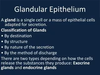
2.Classification of Epithelium 1)Covering epithelium: the epithelium which cover body surface or line the inner surface of body cavities, tubes and sac. 2)Glandular epithelium: the epithelium which main function is secretion. 3)Sensory epithelium: the epithelium which has special sensory function.