Download

1 / 64
640 likes | 710 Views
SIMPLE EPITHELIUM. Dr Mukesh Singla. Epithelial Tissue -- General Features. Closely packed cells with little extracellular material Many cell junctions often provide secure attachment. Cells sit on basement membrane Apical (upper) free surface Basal surface against basement membrane

E N D
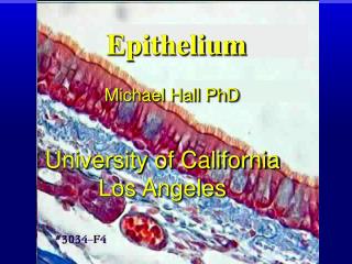
SIMPLE EPITHELIUM Dr MukeshSingla
Epithelial Tissue -- General Features • Closely packed cells with little extracellular material • Many cell junctions often provide secure attachment. • Cells sit on basement membrane • Apical (upper) free surface • Basal surface against basement membrane • Avascular---without blood vessels • nutrients and waste must move by diffusion
Epithelial Tissue -- General Features • Good nerve supply • Rapid cell division (high mitotic rate) • Functions • protection, filtration, lubrication, secretion, digestion, absorption, transportation, excretion, sensory reception, and reproduction.
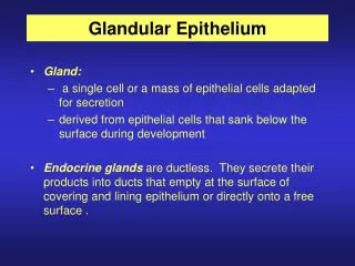
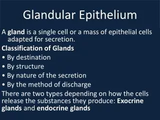
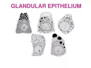
Types of Epithelium 1. Covering and lining epithelium • epidermis of skin • lining of blood vessels and ducts • lining respiratory, reproductive, urinary & GI tract 2. Glandular epithelium- originate from invaginated epithelial cells • secreting portion of glands • thyroid, adrenal, and sweat glands
Epithelium • Epithelium is derived from all three germ layers • Ectoderm-oral and nasal mucosa,cornea , epidermis, glands of skin, mammary glands • Endoderm-Lining of respiratory and gastrointestinal tract, liver , panceas • Mesoderm-lining of urogenital system, circulatory system and body cavities lining-mesothelium
Typical Arrangement of Epithelial Tissue and its Basement Membrane
Basement Membrane • The basement membrane is a thin sheet of fibers that underlies the epithelium • The basement membrane is the fusion of two lamina, the basal lamina-elaborated by epithelial cells and the reticular lamina (or lamina reticularis)-manufactured by cells of connective tissue
Structure of Basement membrane • Basement Membrane • Basal Lamina • Lamina Lucida • Extracellular glycoprotein-Laminin,integrins,entactins,dystroglycans • Transmembrane laminin receptors-project from epithelial cell membrane into basal lamina • Lamina Densa consists of a network of fine filaments. • Type IV collagen. forms felt-like network of fibers that gives the basement membrane its tensile strength
Structure of Basement membrane • Lamina Reticularis • Type III collagen (as reticular fibers) • Attaching proteins (between Basal and Reticular Laminae)-all elaborated by fibroblast of connective tissue • Type VII collagen (anchoring fibrils) • fibrillin (microfibrils) • Fibronectin
lamina lucida &lamina densa • Lamina Densa • dense layer closer to the connective tissue • 30–70 nm in thickness • consists of an underlying network of reticular collagen (type IV) fibrils • Lamina Lucida • clear layer close to epithelium
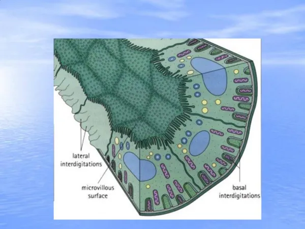
Apical surface Lateral surface Epithelium Basal lamina Reticular lamina Connective tissue
Functions of basement membrane • anchor down the epithelium to its loose connective tissue (the dermis) underneath • provide structural support to the tissue • a mechanical barrier, preventing malignant cells from invading the deeper tissues
Fusion of basal laminae • Glomerularfiltration of the kidney • by the fusion of the basal lamina from the endothelium of glomerular capillaries and the basal lamina of the epithelium of the Bowman's capsule • Gaseous exchange between lungalveoli and pulmonary capillaries • by the fusion of the basal lamina of the lung alveoli and of the basal lamina of the lung capillaries
Cancer cells (Malignant) • If the epithelial cells become transformed (cancerous) and become 'malignant', they are able to break through the basement membrane and invade the tissues beneath. This characteristic is used in the diagnosis of malignant epithelial tumors
A poorly functioning basement membraneDiseases • Genetic defects • Injuries by the body's own immune system • Other mechanisms • Alport syndrome • Genetic defects • Goodpasture's syndrome • Collagen type IV is autoantigen (target antigen) of autoantibodies in the autoimmune disease • Epidermolysisbullosa • Skin • Muscular dystrophy • Dystrophin . a glycoprotein in the plasma membrane of muscle cells re In muscular dystrophy, this protein is defective or missing
Classification Of epithelium According to number of cell layers between basal lamina and free surface and by morphology of epithelial cells • Simple epithelium- composed of single layer of cells • Stratified epithelium- composed of more than one cells
Terms that help us understand what kinds of tissues we are identifying: Terms referring to the layers Simple = one layer Stratified = more than one layer Pseudostratified = false layered (appears to be more than one layer, but only one); ciliated = with cilia Terms referring to the cell shapes Squamous = flat Cuboidal = cube Columnar = rectangular (column) Transitional = ability to change shape
Apical surface Basal surface Simple Apical surface Basal surface Stratified Classification based on number of cell layers.
Squamous Cuboidal Columnar Classification based on cell shape.
The following types of epithelial tissues are covered • in this activity: • 1. Simple squamous epithelial tissue (lungs) • 2. Simple cuboidal epithelial tissue (kidneys) • 3. Simple columnar epithelial tissue (small intestine) • Pseudostratified (ciliated) columnar • epithelial tissue (trachea lining)
The following types of epithelial tissues are covered in this activity
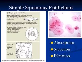
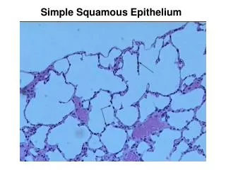
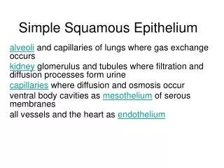
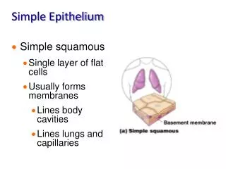
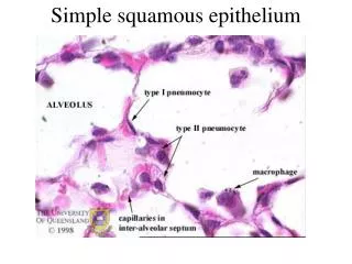
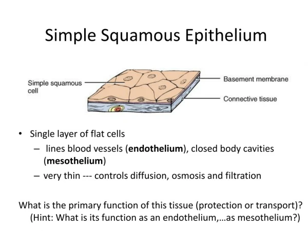
Figure 4.3a Epithelial tissues. (a) Simple squamous epithelium Description: Single layer of flattened cells with disc-shaped central nuclei and sparse cytoplasm; the simplest of the epithelia. Air sacs of lung tissue Function: Allows passage of materials by diffusion and filtration in sites where protection is not important; secretes lubricating substances in serosae. Nuclei of squamous epithelial cells Location: Kidney glomeruli; air sacs of lungs; lining of heart, blood vessels, and lymphatic vessels; lining of ventral body cavity (serosae). Photomicrograph: Simple squamous epithelium forming part of the alveolar (air sac) walls (125x).
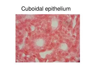
Figure 4.3b Epithelial tissues. (b) Simple cuboidal epithelium Description: Single layer of cubelike cells with large, spherical central nuclei. Simple cuboidal epithelial cells Function: Secretion and absorption. Basement membrane Location: Kidney tubules; ducts and secretory portions of small glands; ovary surface. Connective tissue Photomicrograph: Simple cuboidal epithelium in kidney tubules (430x).
Figure 4.3c Epithelial tissues. (c) Simple columnar epithelium Description: Single layer of tall cells with round to oval nuclei; some cells bear cilia; layer may contain mucus- secreting unicellular glands (goblet cells). Simple columnar epithelial cell Function: Absorption; secretion of mucus, enzymes, and other substances; ciliated type propels mucus (or reproductive cells) by ciliary action. Location: Nonciliated type lines most of the digestive tract (stomach to anal canal), gallbladder, and excretory ducts of some glands; ciliated variety lines small bronchi, uterine tubes, and some regions of the uterus. Basement membrane Photomicrograph: Simple columnar epithelium of the stomach mucosa (860X).
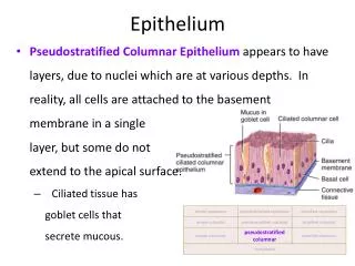
Figure 4.3d Epithelial tissues. (d) Pseudostratified columnar epithelium Description: Single layer of cells of differing heights, some not reaching the free surface; nuclei seen at different levels; may contain mucus- secreting cells and bear cilia. Cilia Mucus of mucous cell Pseudo- stratified epithelial layer Function: Secretion, particularly of mucus; propulsion of mucus by ciliary action. Location: Nonciliated type in male’s sperm-carrying ducts and ducts of large glands; ciliated variety lines the trachea, most of the upper respiratory tract. Basement membrane Photomicrograph: Pseudostratified ciliated columnar epithelium lining the human trachea (570x). Trachea
Given the previous examples(consider the morphology only) Can you name? First, the tissue type Second, where in the body the tissue is found
What kind of tissue does this represent? Simple squamous epithelial tissue Where in the body would you find this tissue? lungs
What kind of tissue does this represent? Simple squamous epithelial tissue (superior view)
What kind of tissue does this represent? Simple cuboidal epithelial tissue Where in the body would you find this tissue? Kidneys (tubules) The lining of the kidney glomerulus (sing.)/glomeruli (pl.) is simple squamous epithelial tissue
What kind of tissue does this represent? Simple columnar epithelial tissue Where in the body would you find this tissue? small intestine
What kind of tissue does this represent? Pseudostratified (ciliated) columnar epithelial tissue “false layered”; it looks like more than one layer, but it is not Where in the body would you find this tissue? trachea lining
What kind of tissue does this represent? Stratified squamous epithelial tissue Where in the body would you find this tissue? mouth lining
What kind of tissue does this represent? Stratified cuboidal epithelial tissue Where in the body would you find this tissue? salivary glands, sweat glands
What kind of tissue does this represent? Stratified columnar epithelial tissue Where in the body would you find this tissue? male reproductive tract
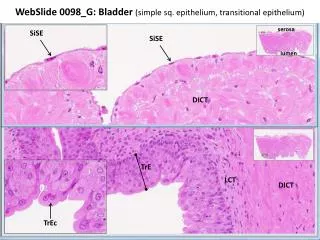
What kind of tissue does this represent? Transitional epithelial tissue Where in the body would you find this tissue? empty bladder
What kind of tissue does this represent? Transitional epithelial tissue Where in the body would you find this tissue? distended (full) bladder
Figure 4.3e Epithelial tissues. (e) Stratified squamous epithelium Description: Thick membrane composed of several cell layers; basal cells are cuboidal or columnar and metabolically active; surface cells are flattened (squamous); in the keratinized type, the surface cells are full of keratin and dead; basal cells are active in mitosis and produce the cells of the more superficial layers. Stratified squamous epithelium Function: Protects underlying tissues in areas subjected to abrasion. Nuclei Location: Nonkeratinized type forms the moist linings of the esophagus, mouth, and vagina; keratinized variety forms the epidermis of the skin, a dry membrane. Basement membrane Connective tissue Photomicrograph: Stratified squamous epithelium lining the esophagus (285x).
Figure 4.3f Epithelial tissues. (f) Transitional epithelium Description: Resembles both stratified squamous and stratified cuboidal; basal cells cuboidal or columnar; surface cells dome shaped or squamouslike, depending on degree of organ stretch. Transitional epithelium Function: Stretches readily and permits distension of urinary organ by contained urine. Location: Lines the ureters, urinary bladder, and part of the urethra. Basement membrane Connective tissue Photomicrograph: Transitional epithelium lining the urinary bladder, relaxed state (360X); note the bulbous, or rounded, appearance of the cells at the surface; these cells flatten and become elongated when the bladder is filled with urine.
Cell junctions • Cell junctions consist of multiprotein complexes that provide contact between neighboring cells or between a cell and the extracellular matrix. • They also build up the paracellular barrier of epithelia and control the paracellular transport. • Cell junctions are especially abundant in epithelial tissues.
Cell to Cell Junctions and Adhesion • A. Cell Adhesion Molecules • B. Cell-Cell Junctions • 1. Occluding Jxs- zonula occludens or tight junctions • 2. Anchoring Jxs • a. Desmosomes or macula adherens(adhesive spots) • b. Zonula adherens or adhesive belt • c. Fascia adherens or adhesive strips • d. Hemidesmosomes • 3. Communicating Jxs or gap junctions
Formation of multicell organisms requires specific interaction • between cells to hold the cells together and to communicate • in order to coordinate activities. • A. 4 types of Cell Adhesion Molecules (CAMs) • are used to hold animal cells together: • Cadherins • Ig-like CAMs • Selectins • Integrins • All are single-pass transmembrane proteins anchored to the • cytoskeleton by their cytoplasmic domains.
Importance of Cell junction • Cell junctions enable communication between neighboring cells via specialized proteins called communicating junctions. • Cell junctions are also important in reducing stress placed upon cells. • Combined with CAMs( cell adhesion molecule) and ECM, cell junctions help hold animal cells together.
Cell junction molecules Four main types: • Selectins, • Cadherins • Integrins • Immunoglobulin superfamily