Download

1 / 29
300 likes | 636 Views
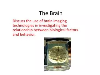
Biological Bases of Behaviour. Lecture 7: Techniques for Understanding Brain Structure & Function. Kalat (2000) p72. Learning Outcomes. At the end of this lecture you should be able to: 1 . Describe a range of techniques used to determine brain structure and function.

E N D
Biological Bases of Behaviour.Lecture 7: Techniques for Understanding Brain Structure & Function. Kalat (2000) p72
Learning Outcomes. • At the end of this lecture you should be able to: • 1. Describe a range of techniques used to determine brain structure and function. • 2. List the advantages and disadvantages of each of the techniques described.
The Techniques • Many techniques have been developed to enable biological psychologists to understand how the brain works, each having their own advantages and disadvantages. • It is rare that a single method will provide a convincing explanation, more often one or more techniques are utilised to provide a clearer picture. • This is referred to as 'converging operations' (Carlson, 1994).
1. Neuroanatomical Techniques. • These tell us about the anatomical structure of the brain. • a) Histological Procedures: Gross examination of the brain does not allow us to study details of cell structure and connectivity, to do so we need to selectively stain thin slices of the brain. • Preservation: After death the soft brain tissue is destroyed by autolytic enzymes, so the brain must be preserved with a fixative, such as formalin. • The brain is then embedded within a paraffin block that can be sliced thinly using a microtome and mounted on slides. • Histological stains have been developed so that cell bodies, nerve fibres and membranes can be selectively viewed
Staining. • 3 types are used: • i) Cell-body stains. • Developed by Franz Nissl who discovered that dyes such as cresyl violet would selectively reveal the cell bodies of brain tissue. • ii) Myelin stains. • These selectively colour the myelin sheath that surrounds nerve cells and so fibre bundles are revealed. Carlson (1994) p 110
Membrane Stains • These contain salts of various heavy metals that interact with the axon membranes. • The commonly-used Golgi-Cox stain uses silver. • This enables us to see the branching of individual neurons and trace their connections. Carlson (1994) p 111
b) Tracing Connections. • The structures of the CNS are interconnected by complex systems of axons, finding out what nuclei are connected to what others and the routes taken can be solved by: • i) Anterograde (forward) tracing: Certain proteins are taken up by cell bodies are transported through axons until they reach the terminal buttons. Lectins such as phaseolus vulgaris leukoagglutinin (PHA-L) are often used. • ii) Retrograde (backward) tracing: Dyes such as flurogold are injected into the terminal buttons and are then carried back through the axons to the cell bodies where they can be seen.
PHA-L Anterograde Tracing. PHA-L injected and taken up by dendrites and cell bodies Transported to terminal buttons PHA-L labelled axons and terminal buttons can be seen under the microscope Carlson (1994) p 111
c) Histochemical Techniques. • These tell us the location of specific neurons that produce and secrete particular neurotransmitters. • Antibodies for a specific neurotransmitterare injected into a region and the slides are viewed under ultraviolet light. • Alternatively radioactive 2-deoxyglucose (2-DG) is taken up by active neurons. Vasopressin-containing axons and terminal buttons Carlson (1994) p 114
2. Imaging the Living Brain. • The methods previously described have all required the brain to be removed. • The following techniques enable neural structure and function to be viewed in the living brain. • Computerised Axial Tomography (CAT): Consists of a circular arrangement of x-ray emitters and detectors in which progressive scans through the brain can be taken. • A 2-dimensional image of horizontal sections can then be produced. • This technique is used mainly to diagnose neurological conditions such as tumours, blood clots, degenerative disease and the location of strokes.
CAT scan from a patient with a lesion in the right occipital-parietal area. Lesion site Carlson (1994) p 119
Magnetic Resonance Imaging (MRI). • MRI takes detailed pictures, using a strong magnetic field. • It detects radiation from hydrogen molecules present in all brain tissue in different concentrations. • Sagittal, horizontal and frontal images are produced. • MRI has now been adapted to show function as well - this is called functional magnetic resonance imaging (fMRI). Carlson (1994) p 120
Positron Emission Tomography (PET). • Radioactive glucose is taken up by active cells in the brain. • As the radioactive isotopes decay, they emit positrons which are detected by the scanner. • In a typical experiment, images of blood flow or radioactive counts during a control state are subtracted from images taken during functional activation (i.e. when the individual is performing some type of cognitive task). • By subtracting measurements in the control state from a task state it is possible to identify those areas of the brain concerned with specific mental operations (Raichle, 1994).
PET Scan of Brain Activation. Alzheimer’s patient Normal elderly control Rosenzweig et al (2002) p 53
Advantages/Disadvantages of Brain Imaging. • Advantages. • Non-invasive (CAT, MRI). • Provide very detailed knowledge about structure (CAT, MRI) and function (PET, fMRI). • Disadvantages. • Mildly invasive (PET) • Only provide horizontal pictures (CAT)
3. Recording Electrical Activity in the Brain. • Axons generate action potentials, and terminal buttons elicit postsynaptic potentials. • These electrical events can be recorded, and changes in electrical activity can be used to determine whether a structure or region of the brain is involved in a certain behaviour. • There are two types of measure: • a) Microelectrodes: Are very small and can record electrical activity within single neurons (single-cell recording). • These are normally implanted chronically into the brain of an animal thus allowing the monitoring of activity as the animal responds to particular environmental stimuli.
Advantages/Disadvantages of Microelectrodes. • Advantages. • Extremely precise. • Disadvantages. • Time consuming. • Too focused - it neglects neuronal interactions. • Invasive.
b) Macroelectrodes. • The Electroencephalogram (EEG) was invented by Berger (1929). • Electrodes are attached to the scalp and the activity of hundreds of thousands of neurons in the vicinity of the electrodes recorded. • Active electrodes are placed over the site of expected neural activity and an indifferentelectrode is placed at a neutral spot (usually the earlobe). • The recording simply measures the potential difference between the two electrodes. • In clinical studies, many electrodes are used and they are placed over the lobes of the brain according to a conventional scheme.
The EEG Record. • Changes in electrical activity are evident in states such as sleep, wakefulness, and arousal;abnormal electrical activity can signal epilepsy or mental illness. • Each individuals EEG pattern is distinctive, but there are characteristic patterns of electrical activity: • Alpha waves (8-13 Hz): Associated with relaxed wakefulness • Beta waves (13-30 Hz): Seen in individuals who are awake, alert, with eyes open, and who may be concentrating on something. • Delta waves (0.5-4 Hz): Associated sleep in adults but are also seen in infants, their abnormal appearance in awake adults can be indicative of a brain tumour. • Theta waves (4-7 Hz): Also seen in adults sleeping and in children. Their abnormal appearance in adults is typically seen in psychopaths.
Advantages/Disadvantages of Macroelectrodes. • Advantages. • Non-invasive • Can differentiate between different neurological conditions or behavioural states. • Disadvantages. • Time consuming. • Very crude - the averaging of activity in many neurons cannot establish precise activity in a particular region.
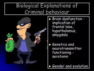
4. Brain Stimulation. • In animals, direct electrical stimulation of the brain can produce clear behavioural changes. • E.g stimulation of the hypothalamus may produce feeding, drinking, sexual arousal, and aggression suggesting an activational role. • Stimulation of the caudate nucleus often halts ongoing behaviour which suggests an inhibitory role. • In humans this technique was pioneered by Penfield and Jasper (1954) in which they stimulated various regions of cortex in conscious patients and noted down the behavioural or sensory effects.
Electrical Stimulation of the Human Brain Carlson (1994) p 137
Advantages/Disadvantages of Brain Stimulation. • Advantages. • Does not harm the brain. • A valid way of investigating living function of brain areas. • Disadvantages. • Invasive. • Crude - not easy to tell how far the stimulation has spread.
5. Experimental Brain Damage. • An influential, though ethically controversial technique is to cause localised brain damage in animals and note the behavioural effects. • If an animal no longer performs a specific behaviour following brain damage to a particular area (a lesion) then that area must be responsible for the behaviour. • There are several methods: • Aspiration (ablation): the surface of the cortex is removed. • Radiofrequency lesion: an electrode is inserted to the correct location and then the tip is heated destroying nearby cells. • Neurotoxic lesion: a neurotoxin such as ibotenic acid is injected into a specific region, this destroys cell bodies but leaves undamaged fibres of passage.
A Radiofrequency Lesion. Bilateral radiofrequency lesion of the cingulum bundle in a rat brain
Problems in Interpreting Results of Brain Lesions in Animals. • 1. How do we know that the damage that has been caused has actually interfered with the behaviour in question? • 2. All regions of the brain are interconnected at some level and so by damaging a structure, we may also damage fibre pathways between other areas. • 3. As the brain works as an integrated whole, disturbance at one location may affect the functioning of other regions. • 4. Following damage, some form of regeneration may take place or other brain regions may partly take over. • 5. We are generalising from a damaged animal to functioning in normal animals.
6. Human Cases of Brain Damage. • Head injuries occur in many different forms but rarely produce localised damage. • Case studies of individuals who do have circumscribed damage can shed light on how the brain functions. • Most cases show a dissociation of impairments, comparisons can be made of the brain regions damaged in different cases. • While it is unethical to administer selective neurotoxins to humans and then observe the effects, some willingly self-administer neurotoxic substances such as ecstasy and provide ready-made experiments on the effects of brain damage on mood, memory, and behaviour.
Advantages/Disadvantages of Brain Damage. • Advantages. • No ethical problems as the damage has occurred naturally. • Disadvantages. • Lack of precision - extent of the damage is not controllable. • There are problems with comparison - ie if a person suffers brain damage and behaves aggressively how do we know that this is not how they behaved before?
References and Bibliography. • Carlson, N.R. (1994). Physiology of Behaviour. • Kalat, J.W. (2000). Biological Psychology. • Penfield, W., & Jasper, H. (1954). Epilepsy and the functional anatomy of the human brain. Boston: Little, Brown & Co. • Raichle, M.E. (1994). Imaging the mind: studies with modern imaging techniques. Annual Review of Psychology, 45: 333 - 356. • Rosenzweig, M.R., Breedlove, S.M., & Leiman, A.L. (2002). Biological Psychology. • Toates, F. (2001). Biological Psychology.