Download
1 / 29
290 likes | 606 Views
Biological Bases of Behaviour. Lecture 8: Central Nervous System. Learning Outcomes. At the end of this lecture you should be able to: 1 . Understand key anatomical terms. 2 . Describe the gross organisation of the CNS. 3 . Identify key structures of the CNS.

E N D
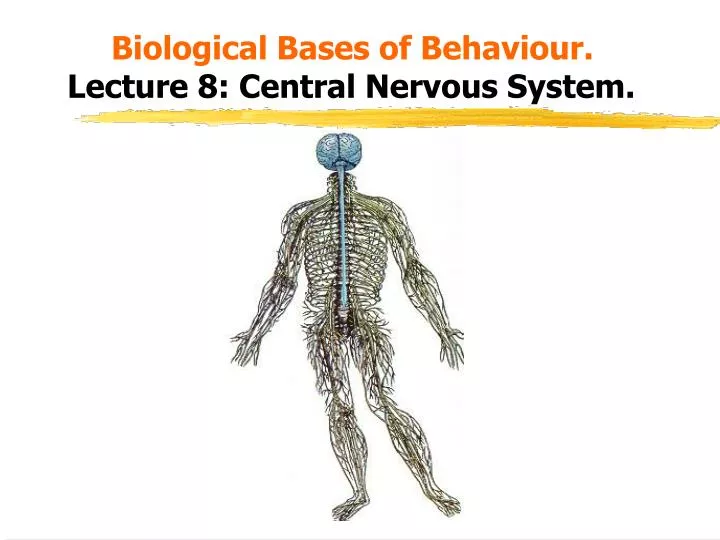
Biological Bases of Behaviour.Lecture 8: Central Nervous System.
Learning Outcomes. • At the end of this lecture you should be able to: • 1. Understand key anatomical terms. • 2. Describe the gross organisation of the CNS. • 3. Identify key structures of the CNS. • 4. Briefly describe the functions of key CNS structures.
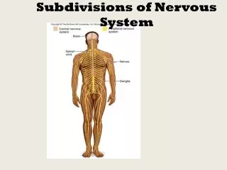
Divisions of the Nervous System. NERVOUS SYSTEM Peripheral Nervous System Central Nervous System Autonomic Nervous System Somatic Nervous System Spinal cord Brain Forebrain Hindbrain Midbrain
Terms of Reference. • When describing the CNS we use directional terms described relative to an imaginary line drawn through the spinal cord and the front of the brain (neuraxis). • Anterior (or rostral): front part of a structure. • Posterior (or caudal): back part of a structure. • Dorsal: Pointing out of the back. • Ventral: Pointing towards the ground. • Lateral: towards the sides. • Medial: towards the middle. • Ipsilateral: on the same side of the body or brain. • Contralateral: on the opposite side of the brain or body.
Anatomical Planes. • Horizontal: a slice parallel to the ground. • Sagittal: a slice perpendicular to the ground, parallel to the neuraxis. • Transverse (coronal): a slice vertical from front to back
Blood Supply. • The brain receives 20% of the blood flow from the heart. • The brain cannot store glucose nor can it extract energy without oxygen, so continuous blood flow is essential. • A 1-second interruption in blood flow will use up all of the brains resources, a 6-second gap produces unconsciousness. Permanent brain damage occurs after a few minutes. • Two major sets of arteries serve the brain: • 1. The vertebral arteries (serving the caudal portion) enter at the base of the skull. • 2 The internal carotid arteries (serving the rostral portion) ascend the left and right sides of the neck. • These join to form the basilar artery. • There is overlap between the arterial systems so that if a vessel becomes blocked (a stroke), potential damage is minimised.
Blood Supply Continued.. Basilar artery Middle cerebral artery Internal carotid artery
Blood-Brain Barrier. • Nutrients such as oxygen and glucose are delivered to the brain, and waste products such as carbon dioxide are extracted. • Molecules over a certain size cannot enter the brain, this blood-brain barrier preserves the brain from harmful substances in the blood. • The barrier is relaxed in the area postrema where the chemoreceptor trigger zone (CTZ) responds to toxic chemicals in the blood to initiate the vomit reflex.
Meninges. • The brain and spinal cord are protected by tough protective tissue called the meninges, consisting of three layers: • i) Dura mater: The thick and tough but flexible outer layer. • ii) Arachnoid membrane: The soft and spongy weblike middle. • iii) Pia mater: The lower layer closely attached to the skull and spine. • Between the pia mater and the arachnoid membrane lies the subarachnoid space through which cerebrospinal fluid (CSF) passes. • Inflammation of the meninges in the brain or spine caused by viral or other means is called meningitis.
The Ventricular System. • The brain is encased in a protective bath of CSF, manufactured by the choroid plexus. • The brain contains a series of 4 hollow interconnected chambers called ventricles which are filled with CSF. • The largest are the 2 lateral ventricles. • These are connected to the third ventricle located in the middle of the brain. • This is connected to the fourth ventricle via the cerebral aqueduct. • If the flow of the CSF is interrupted (e.g. by a tumour) then this increases pressure in the ventricles and they will expand producing hydrocephalus.
The Ventricular System. Lateral ventricles Third ventricle Fourth ventricle Cerebral aqueduct
The Central Nervous System. • The CNS is split into 3 sections. Forebrain Midbrain Hindbrain
1. The Forebrain. • This region is split into the 2 major components. • a) The Telencephalon. • The hemispheres are separated by the longitudinal fissure. • They are covered by cortex. • A deep cleft is referred to as a fissure, and a shallow one is called a sulcus, each ridge is called a gyrus. Two thirds of the surface of the cortex is hidden in these grooves. • Because cells predominate in the cortex, the cortex has a grey appearance and is referred to as 'grey matter'. • Beneath the surface of the cortex run axons covered by the myelin sheath which is referred to as 'white matter'.
Telencephalon (continued). • The most prominent features of the cortex are: • Lateral fissures. • Central sulcus. • Longitudinal fissure. • These clear divisions are used to help define the different lobes of the brain. • The surface of the hemispheres is divided into four lobes: frontal, temporal, parietal and occipital. • The central sulcus divides the frontal lobe from the parietal lobe, and the lateral fissure divides the temporal lobe from the frontal and parietal lobes. • The hemispheres are connected by a bundle of verve fibres called the corpus callosum.
The Lobes of the Brain. Longitudinal fissure Central sulcus Lateral fissure
Sagittal View of the Brain. Central sulcus Corpus callosum Cerebellum Pons Medulla
Subcortical Divisions of the Telencephalon. • i) The Limbic system. • The hippocampus: crucial for learning and memory. • The amygdala is important for emotion and motivation. • Other parts of the limbic system include the mamillary bodies and cingulate gyrus. Cingulate gyrus Thalamus Mamillary bodies Amygdala Hippocampus
ii)Basal Ganglia. • A collection of nuclei located in the forebrain under the anterior portion of the lateral ventricles. They are involved in movement. Caudate nucleus Thalamus Putamen Globus pallidus Amygdala
b) The Diencephalon. • This region surrounds the third ventricle and consists of the following structures: • i) Thalamus. • Separate but interconnected nuclei receive information from the sensory systems, and relay this information to sensory processing areas in the cortex. • It is a relay system and can thus influence almost the whole of the brain. • It may also play a role in learning and memory.
Thalamus. Somatosensory cortex Motor cortex Frontal cortex Occipital cortex Thalamic nuclei
Diencephalon continued.. • ii) The Hypothalamus. • Comprises 22 nuclei and the pituitary gland. • These control the autonomic nervous system and the endocrine system. • Key aspects of behaviour are co-ordinated from here including feeding, sex, sleep, temperature regulation, and emotional behaviour.
Hypothalamus and Surrounding Structures. Cingulate gyrus Thalamus Fornix Hypothalamus Mamillary bodies Amygdala Hippocampus
2. The Midbrain. • This consists of two major regions: • a). Tectum: This contains two main structures: • Superior colliculus part of the auditory system • Inferior colliculus part of the visual system. • b). Tegmentum: This includes the rostral portion of the reticular formation, 90 interconnected nuclei involved in sensory processing, sleep, arousal, attention, muscle tone, movement and reflexes. • Two key structure of the tegmentum are the: red nucleus and the substantia nigra which are important components of the motor system.
Tectum. Thalamus Superior colliculus Inferior colliculus Pons Medulla
3.The Hindbrain. • This consists of two main divisions: • a) The Metencephalon: Consists of two main structures: • i) The cerebellum: Receives information from sensory systems, the muscles, and the vestibular system. It co-ordinates this information to produce smooth movements. • Damage to the cerebellum (which occurs in such as cerebral palsy) impairs walking, balance, posture, and skilled-motor activity. • ii) The pons: A large bulge on the brain stem involved in sleep and arousal.
Divisions of the Hindbrain. • b) The Myelencephalon. • This contains one major structure called the medulla oblongata which is the most caudal part of the brain stem and it borders the spinal cord. • It contains part of the reticular formation and contains nuclei that control vital functions such as control of breathing and skeletal muscle tone.
4. Spinal cord. • This is located within the vertebrae of the spinal column and it communicates with the sense organs and muscles below the neck. • It consists of two fibre pathways: • Dorsal roots process sensory information • Ventral roots process motor information.
Summary of CNS organisation. • A convenient way to visualise how the brain is organised is to imagine it as three structures each increasing in the complexity of the functions they subserve. • At the core is the medulla-brainstem which controls basic aspects of behaviour (breathing, swallowing, digestion, urination etc). Alongside is the cerebellum which controls movement and posture. • Wrapped around this core is the limbic system, a primitive region responsible behaviours essential to basic survival - pain, pleasure, fear, eating and sex. • The outer layer of the brain is the cerebral cortex - a thin sheet of neurons dealing with higher-order functions such as perception, planning, problem solving thinking, consciousness etc.