Download
1 / 1
10 likes | 125 Views
The subcellular localisation of a metabolic regulator implicated in diabetes. L. Tully 1 , J. Muller 2 , V. Zammit 2 1 School of Life Sciences, University of Warwick, Gibbet Hill Road, CV4 7AL 2 Warwick Medical School, University of Warwick, Gibbet Hill Road, CV4 7AL.

E N D
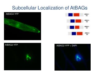
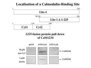
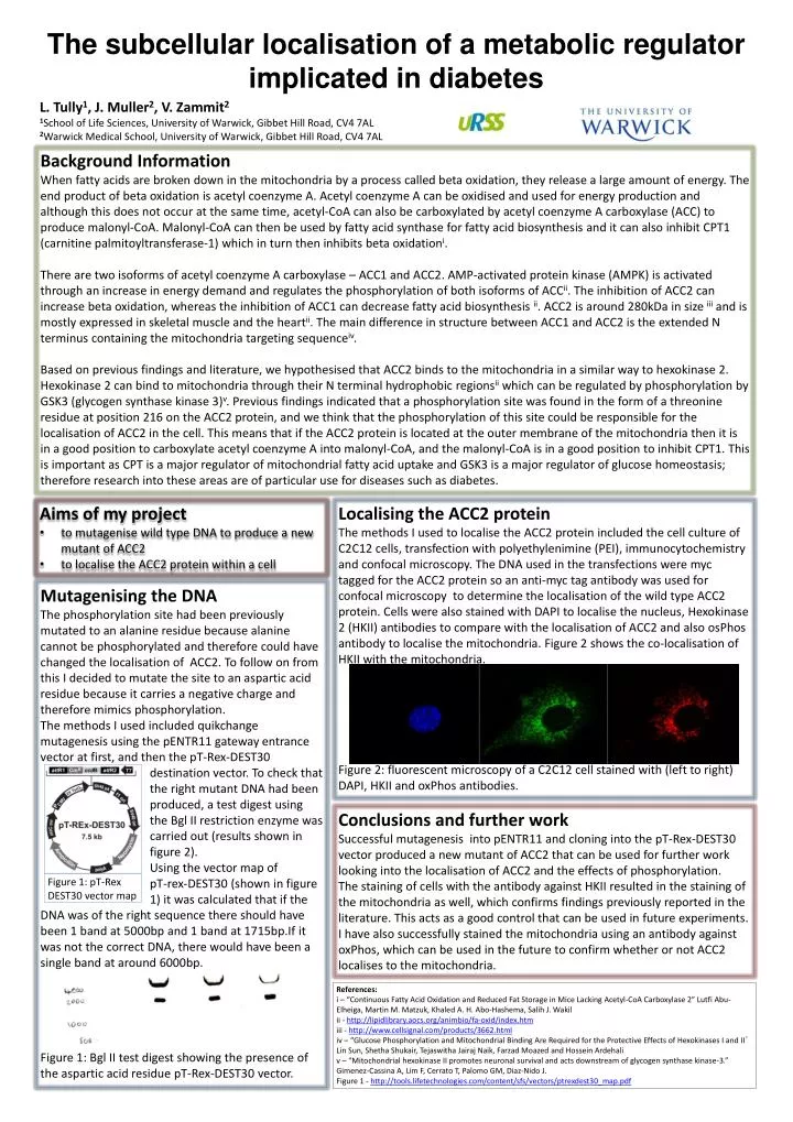
The subcellular localisation of a metabolic regulator implicated in diabetes L. Tully1, J. Muller2, V. Zammit21School of Life Sciences, University of Warwick, Gibbet Hill Road, CV4 7AL2Warwick Medical School, University of Warwick, Gibbet Hill Road, CV4 7AL Background InformationWhen fatty acids are broken down in the mitochondria by a process called beta oxidation, they release a large amount of energy. The end product of beta oxidation is acetyl coenzyme A. Acetyl coenzyme A can be oxidised and used for energy production and although this does not occur at the same time, acetyl-CoA can also be carboxylated by acetyl coenzyme A carboxylase (ACC) to produce malonyl-CoA. Malonyl-CoA can then be used by fatty acid synthase for fatty acid biosynthesis and it can also inhibit CPT1 (carnitine palmitoyltransferase-1) which in turn then inhibits beta oxidationi. There are two isoforms of acetyl coenzyme A carboxylase – ACC1 and ACC2. AMP-activated protein kinase (AMPK) is activated through an increase in energy demand and regulates the phosphorylation of both isoforms of ACCii. The inhibition of ACC2 can increase beta oxidation, whereas the inhibition of ACC1 can decrease fatty acid biosynthesis ii. ACC2 is around 280kDa in size iii and is mostly expressed in skeletal muscle and the heartii. The main difference in structure between ACC1 and ACC2 is the extended N terminus containing the mitochondria targeting sequenceiv. Based on previous findings and literature, we hypothesised that ACC2 binds to the mitochondria in a similar way to hexokinase 2. Hexokinase 2 can bind to mitochondria through their N terminal hydrophobic regionsii which can be regulated by phosphorylation by GSK3 (glycogen synthase kinase 3)v. Previous findings indicated that a phosphorylation site was found in the form of a threonine residue at position 216 on the ACC2 protein, and we think that the phosphorylation of this site could be responsible for the localisation of ACC2 in the cell. This means that if the ACC2 protein is located at the outer membrane of the mitochondria then it is in a good position to carboxylate acetyl coenzyme A into malonyl-CoA, and the malonyl-CoA is in a good position to inhibit CPT1. This is important as CPT is a major regulator of mitochondrial fatty acid uptake and GSK3 is a major regulator of glucose homeostasis; therefore research into these areas are of particular use for diseases such as diabetes. • Aims of my project • to mutagenise wild type DNA to produce a new mutant of ACC2 • to localise the ACC2 protein within a cell Localising the ACC2 protein The methods I used to localise the ACC2 protein included the cell culture of C2C12 cells, transfection with polyethylenimine (PEI), immunocytochemistry and confocal microscopy. The DNA used in the transfections were myc tagged for the ACC2 protein so an anti-myc tag antibody was used for confocal microscopy to determine the localisation of the wild type ACC2 protein. Cells were also stained with DAPI to localise the nucleus, Hexokinase 2 (HKII) antibodies to compare with the localisation of ACC2 and also osPhos antibody to localise the mitochondria. Figure 2 shows the co-localisation of HKII with the mitochondria. Figure 2: fluorescent microscopy of a C2C12 cell stained with (left to right) DAPI, HKII and oxPhos antibodies. Mutagenising the DNAThe phosphorylation site had been previously mutated to an alanine residue because alanine cannot be phosphorylated and therefore could have changed the localisation of ACC2. To follow on from this I decided to mutate the site to an aspartic acid residue because it carries a negative charge and therefore mimics phosphorylation. The methods I used included quikchange mutagenesis using the pENTR11 gateway entrance vectorat first, and then the pT-Rex-DEST30 destination vector. To check that the right mutant DNA had been produced, a test digest using the Bgl II restriction enzyme was carried out (results shown in figure 2). Using the vector map of pT-rex-DEST30 (shown in figure 1) it was calculated that if the DNA was of the right sequence there should have been 1 band at 5000bp and 1 band at 1715bp.If it was not the correct DNA, there would have been a single band at around 6000bp. Figure 1:Bgl II test digest showing the presence of the aspartic acid residue pT-Rex-DEST30 vector. Conclusions and further work Successful mutagenesis into pENTR11 and cloning into the pT-Rex-DEST30 vector produced a new mutant of ACC2 that can be used for further work looking into the localisation of ACC2 and the effects of phosphorylation. The staining of cellswith the antibody against HKII resulted in the staining of the mitochondria as well, which confirms findings previously reported in the literature. This acts as a good control that can be used in future experiments. I have also successfully stained the mitochondria using an antibody against oxPhos, which can be used in the future to confirm whether or not ACC2 localises to the mitochondria. Figure 1: pT-Rex DEST30 vector map References: i – “Continuous Fatty Acid Oxidation and Reduced Fat Storage in Mice Lacking Acetyl-CoA Carboxylase 2”Lutfi Abu-Elheiga,Martin M. Matzuk,KhaledA. H. Abo-Hashema,SalihJ. Wakil ii - http://lipidlibrary.aocs.org/animbio/fa-oxid/index.htm iii - http://www.cellsignal.com/products/3662.html iv – “Glucose Phosphorylation and Mitochondrial Binding Are Required for the Protective Effects of Hexokinases I and II” Lin Sun, Shetha Shukair, TejaswithaJairaj Naik, FarzadMoazed and Hossein Ardehali v – “Mitochondrial hexokinase II promotes neuronal survival and acts downstream of glycogen synthase kinase-3.” Gimenez-Cassina A, Lim F, Cerrato T, Palomo GM, Diaz-Nido J. Figure 1 - http://tools.lifetechnologies.com/content/sfs/vectors/ptrexdest30_map.pdf