Download
1 / 1
10 likes | 123 Views
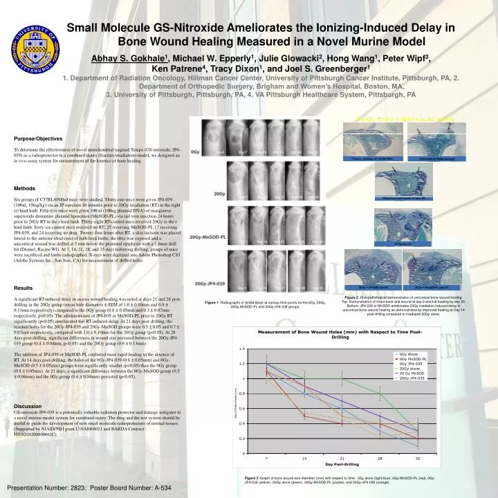
Small Molecule GS-Nitroxide Ameliorates the Ionizing-Induced Delay in Bone Wound Healing Measured in a Novel Murine Model Abhay S. Gokhale 1 , Michael W. Epperly 1 , Julie Glowacki 2 , Hong Wang 1 , Peter Wipf 3 , Ken Patrene 4 , Tracy Dixon 1 , and Joel S. Greenberger 1

E N D
Small Molecule GS-Nitroxide Ameliorates the Ionizing-Induced Delay in Bone Wound Healing Measured in a Novel Murine Model Abhay S. Gokhale1, Michael W. Epperly1, Julie Glowacki2, Hong Wang1, Peter Wipf3, Ken Patrene4, Tracy Dixon1, and Joel S. Greenberger1 1. Department of Radiation Oncology, Hillman Cancer Center, University of Pittsburgh Cancer Institute, Pittsburgh, PA, 2. Department of Orthopedic Surgery, Brigham and Women’s Hospital, Boston, MA, 3. University of Pittsburgh, Pittsburgh, PA, 4. VA Pittsburgh Healthcare System, Pittsburgh, PA Purpose/Objectives To determine the effectiveness of novel mitochondrial targeted Tempo (GS-nitroxide, JP4-039) as a radioprotector in a combined injury (fracture/irradiation) model, we designed an in vivo assay system for measurement of the kinetics of bone healing. Methods Six groups of C57BL/6NHsd mice were studied. Thirty-one mice were given JP4-039 (100ul; 10mg/kg) via an IP injection 10 minutes prior to 20Gy irradiation (RT) to the right (r) hind limb. Fifty-five mice were given 100 ul (100ug plasmid DNA) of manganese superoxide dismutase plasmid liposomes (MnSOD-PL) via tail vein injection 24 hours prior to 20Gy RT to the r hind limb. Thirty-eight RT-control mice received 20Gy to the r hind limb. Sixty-six control mice received no RT; 25 receiving MnSOD-PL 17 receiving JP4-039, and 24 receiving no drug. Twenty-four hours after RT, a skin incision was placed lateral to the anterior tibial crest of both hind limbs; the tibia was exposed and a unicortical wound was drilled at 5 mm below the proximal epiphysis with a 1.6mm drill bit (Dremel, Racine WI). At 7, 14, 21, 28, and 35 days following drilling, groups of mice were sacrificed and limbs radiographed. X-rays were digitized into Adobe Photoshop CS3 (Adobe Systems Inc., San Jose, CA) for measurement of drilled holes. Results A significant RT-induced delay in osseus wound healing was noted at days 21 and 28 post-drilling in the 20Gy group (mean hole diameters ± SEM of 1.0 ± 0.10mm and 0.8 ± 0.13mm respectively) compared to the 0Gy group (0.6 ± 0.05mm and 0.3 ± 0.07mm respectively, p<0.05). The administration of JP4-039 or MnSOD-PL prior to 20Gy RT significantly (p<0.05) ameliorated this RT-induced delay. At 21 days post-drilling, the residual holes for the 20Gy-JP4-039 and 20Gy-MnSOD groups were 0.5 ± 0.05 and 0.7 ± 0.07mm respectively, compared with 1.0 ± 0.10mm for the 20Gy group (p<0.05). At 28 days post-drilling, significant differences in wound size persisted between the 20Gy-JP4-039 group (0.4 ± 0.04mm, p<0.05) and the 20Gy group (0.8 ± 0.13mm). The addition of JP4-039 or MnSOD-PL conferred more rapid healing in the absence of RT. At 14 days post-drilling, the holes of the 0Gy-JP4-039 (0.6 ± 0.05mm) and 0Gy-MnSOD (0.5 ± 0.05mm) groups were significantly smaller (p<0.05) than the 0Gy group (0.8 ± 0.05mm). At 21 days, a significant difference between the 0Gy-MnSOD group (0.5 ± 0.04mm) and the 0Gy group (0.6 ± 0.04mm) persisted (p<0.05). Discussion GS-nitroxide JP4-039 is a potentially valuable radiation protector and damage mitigator in a novel murine model system for combined injury. The drug and the test system should be useful to guide the development of new small molecule radioprotectors of normal tissues. (Supported by NIAID/NIH grant U19AI068021 and BARDA Contract HHSO10200800062C) Figure 2: Histopathological demonstration of unicortical bone wound healing. Top: Demonstration of intact bone and wound at day 0 and full healing by day 35. Bottom: JP4-039 or MnSOD ameliorates 20Gy irradiation induced delay in unicortical bone wound healing as demonstrated by improved healing at Day 14 post-drilling compared to irradiated 20Gy alone. Figure 1: Radiographs of drilled tibiae at various time points for the 0Gy, 20Gy, 20Gy MnSOD-PL and 20Gy-JP4-039 groups. Size of Drilled Holes (mm) PS1.30 Figure 3: Graph of bone wound size diameter (mm) with respect to time. 0Gy alone (light blue), 0Gy-MnSOD-PL (red), 0Gy-JP4-039 (yellow), 20Gy alone (green), 20Gy-MnSOD-PL (purple), and 20Gy-JP4-039 (orange). Presentation Number: 2823; Poster Board Number: A-534