Download
1 / 11
120 likes | 386 Views
Hip Arthroscopy. Mazloumi MD. Introduction. Although hip arthroscopy was first described in 1931, it has only recently become a commonly performed procedure. Pathoanatomy.

E N D
Hip Arthroscopy Mazloumi MD
Introduction • Although hip arthroscopy was first described in 1931, it has only recently become a commonly performed procedure.
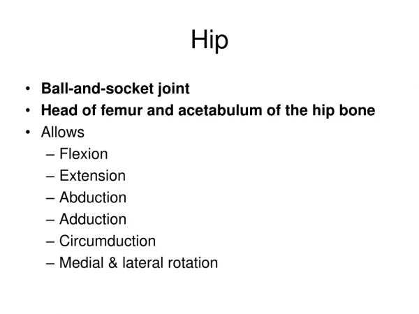
Pathoanatomy • The femoroacetabular joint is a ball-and-socket joint with inherent bony stability, reinforced by a thick capsuloligamentous layer. • The acetabular labrum extends from the anterosuperior to posterosuperior aspects of the acetabular rim
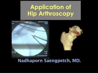
Biomechanic Current indications for hip arthroscopy include • acetabularlabral tear • chondral lesion • loose body removal • infection • synovitis. • femoroacetabular impingement.
Biomechanics Anterosuperiorlabral tear Femoroacetabular impingement (FAI)
Biomechanics • Tears of the acetabular labrum can be the result of athletic injuries at the extremes of motion or from instability events. These injuries can also result in chondral lesions. Femoroacetabular impingement appears to be a developmental anomaly with either retroversion of the acetabulum (pincer lesion), a cam lesion on the femoral head-neck junction, or most commonly, a combination of both (> 50% of cases) A pincer lesion is anterior over-coverage causing the femoral neck to abut against the labrum when in a maximally flexed position. A cam lesion is loss of the normal femoral head-neck offset, or sphericity of the femoral head – which comes into contact with the anterosuperior labrum and acetabulararticular cartilage in the flexed position.
History and Physical positive impingement sign crossover sign
Paraclinics • Magnetic resonance image arthrogram provides excellent visualization of structures such as the ligamentum and labrum and extraarticular soft-tissues. • Diagnostic intraarticular injection may also be performed under fluoroscopic guidance.
Surgical Techniques traditional fracture table anterior aspect of the hip joint
Surgical Techniques Psoastenotomy osteoplasty of the acetabular overhang (“rim trimming”)
Brief Discussion of Results There have been series reporting excellent results with • Loose body removal,6 • Labral debridement,7 • Chondral microfracture,2 • Femoroacetabular decompression.3