Download
1 / 17
170 likes | 259 Views
General Properties of Complex Binding Function - Structure Relationships. Hemoglobin 1. Review of last lecture - myoglobin structure. We noted that the oxygen binding required an unusual symbiosis of an inorganic ion (Fe 2+ )

E N D
General Properties of Complex BindingFunction - Structure Relationships Hemoglobin 1
Review of last lecture - myoglobin structure We noted that the oxygen binding required an unusual symbiosis of an inorganic ion (Fe2+) and a non-proteinaceous hydrophobic group (protoporphyrin IX) and the protein chain. Oxygen binding by Fe2+ is not very strong and even if it were strong would be of not much use as an oxygen carrier since it would diffuse rapidly to regions where it was not needed or be cleared by the kidney. So the Fe2+ ion associates to the protoporphyrin IX - this ultimately becomes the prosthetic group of the protein. Question 1: Why would a Fe2+ ion bind to a heme group? Answer: The Fe2+ has the ability to produce a d2sp3 octahedral hybrid for electron pairs from donors to fill. The heme group can provide 4 electron pairs in this way, leaving two orbitals empty and ready for other donors. However, in aqueous solution the free heme group has very low affinity for oxygen. Question 2: If there are two orbitals left in the hybrid to be filled why does not oxygen bind to these two sites? Answer: It is likely that the oxygen of the water molecule can effectively compete with oxygen to donate electron pairs to the empty Fe2+ orbitals because the molar concentration of water is enormous.
We noted in our last lecture that despite differences in the primary structure, myoglobin and the two hemoglobin chains folded in the same fashion as eight helical segments A - H Therefore, a hydrophobic cavity is present and heme being largely hydrophobic will have a tendency to bind into the pocket. But many proteins have hydrophobic cavities so what is so particular about this one that causes heme to bind with high affinity?
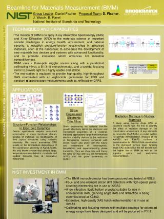
Role of the conserved histidines: The cavities to which heme binds in myoglobin and hemoglobin have two conserved histidines one in the E-helical segment (distal histidine) and one in the F-helix (proximal histidine). Proximal Histidine. One of the N atom of the imidazole ring of the proximal histidine donates a lone pair to the heme anchoring it in the cavity and leaving one remaining empty orbital to be filled. The net effect of the binding into the hydrophobic cavity is the heme is now shielded from the aqueous medium by the protein and oxygen can diffuse and reach it to donate its electron pair. Distal Histidine. This histidine does not form any coordination bond with the heme but it is close enough to prevent the linear oxygen molecule to attach perpendicularly to the plane of the heme. In this way, the oxygen binding is weakened slightly to allow for its displacement when the free oxygen concentration falls to a critical value.
This effect on the oxygen binding by the distal his is not so apparent in the stick and ball model representation shown above.
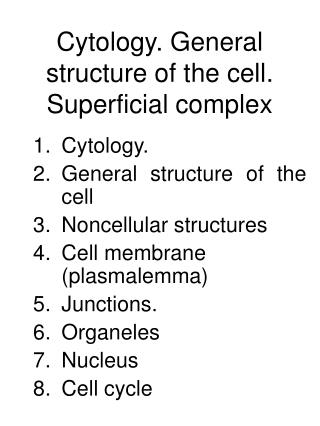
distal his It is more obvious in this space filling representation.
Hemoglobin. Tetrameric protein made of four subunits (chains) a2b2 made up of two ab dimers. The quarternary structure shows that these subunits are arranged tetrahedrally. The molecule is essentially spheroidal (64 x 55 x 50 Å) The binding sites are the four hemes associated with each subunit
From the oxygen binding studies we know that the binding behavior is complex. That is the direct plot of oxygen binding by hemoglobin is sigmoidal and not hyperbolic. This signifies that the sites while identical are not independent, meaning that the binding of the first oxygen facilitates the binding of the next oxygen to the molecule. Normally, if there is going to be dependence between the sites this usually signifies a direct effect implying that the sites are close enough to affect one another. We can examine the structure and measure the distances between the Fe atoms of the hemes by using the DTMM modeler (make sure the auto valency in the toggle menu (style) is unchecked) and then deleting the protein chains leaving the hemes and some water molecules . Then by using the calculate menu we can get the distances.
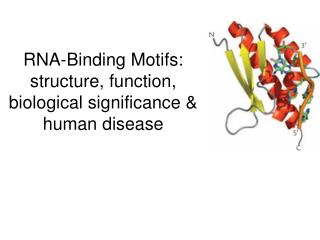
If the hemes are too far apart to influence the binding of one another, then how does this dependency occur? oxy-hemoglobin deoxy-hemoglobin Binding of oxygen produces a change in the position of the F-helix in the subunit and this propogates and this possibly alters the interactions between the other subunits thereby altering their conformations slightly. In some fashion this makes the remaining hemes easier to bind too.
If the hemes are too far apart to influence the binding of one another, then how does this dependency occur? We have earlier examined how we can analyze the binding data using the Hill analysis, but as we saw then, the Hill mechanism is not a valid molecular model. Why not? The mechanism for binding is that c oxygen molecules all bind at the same time or dissociate at the same time: Hb (O2)c = Hb + c O2 Since c does not have to be integral, this is not a valid molecular mechanism but it does provide a way to analyze cooperative binding. The Hill equation and the graphical representation allows for the determination of c, the Hill coefficient, from the slope and the dissociation constant, Kd, for the above mechanism from the y axis intercept. log Y = - c log [B50] + c log [B] 1-Y
There are two molecular models that can explain the cooperative binding effect: 1. The sequential model 2. The concerted model The sequential model for the binding suggests a conformational change in the subunit containing the heme binding the oxygen and as a consequence, this change is propogated to the other subunits which have no oxygen bound to them but these changes increase the affinity for subsequent binding of oxygen to the other sites. Hb + O2 = Hb O2 k1 Hb O2 + O2 = Hb (O2)2 k2 Hb(O2)2 + O2 = Hb (O2)3 k3 Hb(O2)3 + O2 = Hb(O2)4 k4 In this model, the intrinsic binding constants, k’s (while identical initially) change as oxygen loads one at the time to the hemoglobin. Recall, that we have to represent these as dissociation reactions and, therefore, the equilibrium reactions should be shown as the reverse of those given above.
The concerted model is very different in that it can explain the sigmoidal direct curve by assuming that the sites are identical and independent. This seems to be defying what we know about the anticipated behavior for multiple identical and independent sites, since we know that this type of binding gives hyperbolic and not sigmoidal direct plots. If the intrinsic equilibrium dissociation constants are the same and do not change with binding, then how can one get a sigmoidal direct plot? The reason we can get sigmoidal binding behavior is that we make the assumption that the protein exists in two different conformations in equilibrium with one another. Furthermore, we also assume that the ligand (oxygen) binds preferentially only to one form. Ro = To Therefore, L = [To] [Ro] where the subscript o indicates the equilibrium when there is no ligand (in this case oxygen) present. L is called the allosteric constant. We assume that binding can only takes place to the R form and that the binding to the sites in R occurs as independent and identical.
Let the intrinsic dissociation constant for the binding of ligand to any of these sites be kR With this information you must be able to derive the binding function relating Y to the concentration of free ligand [B], the intrinsic dissociation constant, kR, and the allosteric constant L. The function is: Y = a(1 + a) / {(1 + a)2 + L} where a = [B] / kR
Some features of oxygen binding by hemoglobin: 1. Only the ferro-form binds oxygen - the ferri-form sometimes referred to as methemoglobin or metmyoglobin does not bind oxygen. 2. Carbon dioxide does not bind to the heme group - most of it is in the form of dissolved CO2 ( and H2CO3) or in a modified form involved in the carbamylation of the a-NH2 of the chains. 3. Carbon monoxide can bind to ferro-hemoglobin - the binding is strong but because of the distal histidine the CO must bind at an angle and this is thought to lower its affinity somewhat. 4. Oxygen-binding causes a red shift in the spectrum of the bound heme. This spectral change in the visible range can be used to measure the amount of oxygen bound. 5. The binding of oxygen is affected by effectors such as H+ (pH) and this is called the Bohr effect. 6. The binding is also affected by certain small organo phosphate molecules such as 2,3-bisphosphoryl glycerate (BPG)