Download
1 / 53
530 likes | 670 Views
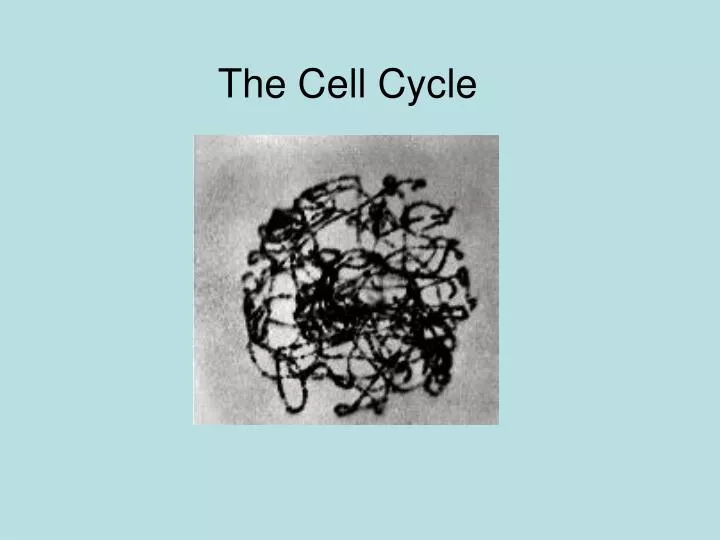
The Cell Cycle. DNA has a Double helix shape. This is a naked strand of DNA. DNA wraps itself around proteins. DNA + protein is called Chromatin. Visible, compact chromatin makes up a Chromosome. As chromatin condenses…. it becomes. visible under a light microscope.

E N D
DNA has a Double helix shape This is a naked strand of DNA DNA wraps itself around proteins DNA + protein is called Chromatin Visible, compact chromatin makes up a Chromosome As chromatin condenses… it becomes visible under a light microscope.
Naked DNA Chromatin Supercoils Chromosome This is another look at how DNA is packaged.
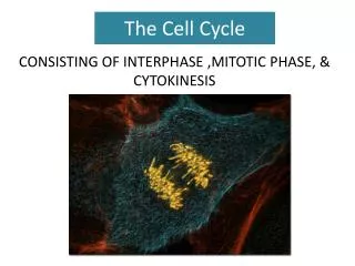
The three C’s that might confuse you. Centrosome Centrioles Centromeres
Centrosomes Nucleolus Nucleus The Cell in Interphase Cell membrane DNA “G1” stage Replicates (“S” stage) Cytoplasm Chromatin forms supercoils (“G2” stage)
Nucleolus disappears Chromosomes become visible The Cell in Early Prophase Chromatin condenses
The Cell in Late Prophase Nuclear envelope breaks down Centrosomes move apart Spindle fibers form
METAPHASE Spindle fibers attach to centromeres Chromosomes align in middle of the cell
METAPHASE Under the microscope, chromosomes may appear flat. In the cell however, chromosomes have a three-dimensional arrangement.
Sister Chromatids separate and move apart ANAPHASE “Free” spindle fibers lengthen; cell elongates Spindle microtubules disintegrate behind chromatids
TELOPHASE Daughter chromosomes reach centrosomes
CYTOKINESIS TELOPHASE Mitotic spindle disappears Cell divides into two identical daughter cells Nuclear membrane reforms Nucleoli reappear
New daughter cells INTERPHASE CYTOKINESIS
Mitosis Animated Click through the next 30 slides to see a rough animation of the cell cycle.
Prophase 8
Prophase 9
Prophase 10
Prophase 11
Metaphase 12
Metaphase 13
Metaphase 14
Anaphase 15
Anaphase 16
Anaphase 17
Anaphase 18
Telophase 19
Telophase 20
Telophase 21 Cytokinesis
Telophase 22 Cytokinesis
Telophase 23 Cytokinesis
Telophase 24 Cytokinesis
Telophase 25 Cytokinesis
Interphase 29
Interphase 30
MITOTIC EVENTS PRACTICE IDENTIFY THE MITOTIC EVENTS IN THE SLIDES WHICH FOLLOW
What mitotic event is shown? Answer: Anaphase
What mitotic event is shown? Answer: Prophase