Download
1 / 36
360 likes | 372 Views
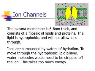
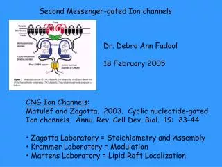
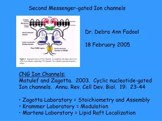
Second-Messenger Gated Ion Channels. Tom Mast Membrane Biophysics 10/5/07. An intracellular signal produced in response to a stimulus usually when a ligand binds a receptor ex: cyclic nucleotides (cAMP or cGMP) calcium inositol 1,4,5 triphosphate (IP3)

E N D
Second-Messenger Gated Ion Channels Tom Mast Membrane Biophysics 10/5/07
An intracellular signal produced in response to a stimulus usually when a ligand binds a receptor ex: cyclic nucleotides (cAMP or cGMP) calcium inositol 1,4,5 triphosphate (IP3) diacylglycerol (DAG) What is a Second Messenger?
Classic Physiological Role of Second Messenger: cAMP in Rods Things to notice: Amplification steps Modulation Change in ion flow Calcium Feedback Ligand binding http://openwetware.org/wiki/BIO254:DarkNoise
Second Messenger Channel Topics Ion species flux- specific or not, calcium Structure- subunit architecture pore and selectivity filter conformational changes tetramerization Ligand-binding- what is the ligand conformation/ shape changes kinetics
Which Channels? Cyclic gated nucleotide Channel A 2 Transient Receptor Potential C 2 Inositol 1,4,5 Triphosphate 1
Common Subunit Structure IP3 R1 Bosanac et al., BBA vol.1742 December 2004, 89-102 http://www.ukbf.fu-berlin.de/pharma/agschaefermulti.html
Channel Pore and Selectivity Sequence Bovine Rod CNGC RKYVYSLYWSTLTLTTIG..ETPPPV Catfish Olfactory CGNC FCYVYCFYWSTLTLTTIG..EMPPPV Bacterial K KSA TYPRALWWSVETATTVGYGDLY.PY Shaker K SIPDAFWWAVVTMTTVGYGDMT.PV Mammalian IP3R1 LLMCIVTVLSHGLRSGGGVGDVLRK Mammalian TRPC2 FNETFQFLFWTMFGMEEHTVVDMP Common Pore Region Other regions within the channels are similar (ie S4) Original channel may have been a 1 TMD Ca++ Strong et al., Mol. Bio. Evol. 1993 (10) 221-242 Due to these relatively non-selective pore regions these channels flux cations mainly Na+ and Ca++
TRPC2 and IP3R1 Signaling C. Badland
The TRP Channels Notice: weak voltage sensor and ‘TRP’ box
Paper 1: TRPC2 Lucas P, Ukhanov K, Leinders-Zufall T, Zufall F A Diacylglycerol-Gated Cation Channel in Vomeronasal Neuron Dendrites Is Impaired in TRPC2 Mutant Mice Neuron. 2003 Oct 30;40(3):551-61. Primary Question: Are DAG-induced currents present in VNO neurons?
Figure 1 A- sensory neuron in vitro B- inside-out patch: response to DAG analogue C- F I-V relationship of SAG-induced currents note: permeable to several ion species outward current block by large cation
Figure 2 inside-out patch: single channel responses to SAG A Low spontaneous opening w/o SAG B. increased opening C-D Frequency histograms of opening E I-V relationship of SAG-induced currents
Figure 3 Ligand specifity of the SAG-induced current Important data: IP3 does not gate current neither do all fatty acids
Figure 4 • Whole-cell currents • w/o SAG in WT • w/ SAG in WT • W/SAG in WT • and bath application • of large cation • D-F. Same a A-C except • in TRPC2 KO • G. I-V relationship WT • H. I-V relationship KO • I. Histogram of SAG • induced currents Important Data: TRPC2 KO neurons Lack the SAG-induced current of WT
Figure 5 • Whole cell recording in Current-clamp • dilute urine activates neuron • C-D. This activation has an I-V relationship • similar to SAG-induced currents
Figures 6 + 7 Below: Phospholipase C inhibitor (U-73122) blocks the urine-induced current in voltage-clamped neurons Above: DAG kinase inhibitor induces an inward Current which is abolshed by Phospholipase C inhibitor (U-73122) in voltage-clamped neurons Important data: in neurons pharmacologically increasing or decreasing endogenous DAG produces the predicted result
Conclusions A urine induced current is non-selective for external cations It is dependent on PLC It is abolished by gene-targeted deletion of TRPC2 It closely resembles that of a SAG-induced current
Paper 2: IP3 R1 Hamada K, Terauchi A, Mikoshiba K. Three-dimensional rearrangements within inositol 1,4,5-trisphosphate receptor by calcium. J Biol Chem. 2003 Dec 26;278(52):52881-9. Primary Question: How do the allosteric factors Ca++ and IP3 effect conformational changes in the channel?
Simple example of Allostery http://biology.fullerton.edu/biol302/regulation.html Binding of a factor at one site alters other sites could be enzymatic activity, affinity, conformation
Figure 1 • A . Incubation of IP3R1 with a • lysine-protease results in • different fragment patterns • dependent on Ca++ concentration • Also dependent on C-term of • cytoplasmic domain which is • involved in tetramerization • C. Location of epitopes used in • western analysis
Figure 2 + 3 Left: Mg++ does not affect proteolysis while Sr++ and (maybe) Ba++ Does. IP3 does not affect proteolysis. Right: IP3R1 favors a ‘windmill’ shape in certain Ca++ concentrations
Figure 6 • Modeling the 3-D • Shape of IP3R1 • Based on • Transmission • Electron micrographs (fig 4) • w/o Ca++ • w/ Ca++
Figure 7 + 8 w/o Ca++ w/ Ca++
Conclusions IP3R1 is sensitive to Ca++ Channel-wide conformational changes are due to Ca++ binding and not IP3 Tetramerization may play a role in the conformational changes
Paper 3: CNGA2 Nache V, Schulz E, Zimmer T, Kusch J, Biskup C, Koopmann R, Hagen V, Benndorf K Activation of olfactory-type cyclic nucleotide-gated channels is highly cooperative J Physiol. 2005 Nov 15;569(Pt 1):91-102 Primary Question: What is the allosteric model for cGMP binding to CNGA2?
Summary of the Canonical Cilia Cascade CNCGs consist of three subunits: A2:A4:β1 in a 2:1:1 ratio A2 is required to detect most odors.
Calculating Cooperative Binding An allosteric relationship Binding of the first ligand changes the affinity for future ligands at other to other sites h > 1 = positive h < 1 = negative H cannot be greater than the # of binding sites
Figure 1 • Experimental set-up: • inside-out patches exposed • to light-sensitive cGMP • B. Light pulse • C. Example current
Figure 2 • Example of current • used for calculations B-D [cXMP]-response curves with calculated parameters
Figure 3 • Activation time-courses • Plot of time-constants • Plot of activation ratios • Voltage-effect
Figure 4 Activation w/ cAMP is slower At a rate consistant W/ binding Indicates changes in activation over [cGMP] is intrinsic
Figure 5 Channels in native Ratios (2:1:1) have Similar activation when Compared to CNGA2 Indicates kinetics are intrinsic
Figure 6 CNGA2 channels open spontaneously has implications as to type of allosteric model
Figure 8 Activation kinetics are best fit by model with 3 binding steps and with both negative and positive cooperativity
Conclusions CNGA2 channels have a greater affinity for cGMP CNGA2 channels display cooperative binding CNGA2 and hetereomultimer channels are affected by Vm