Download
1 / 18
180 likes | 317 Views
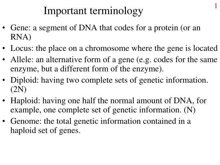
Important terminology. Gene: a segment of DNA that codes for a protein (or an RNA) Locus: the place on a chromosome where the gene is located Allele: an alternative form of a gene (e.g. codes for the same enzyme, but a different form of the enzyme).

E N D
Important terminology • Gene: a segment of DNA that codes for a protein (or an RNA) • Locus: the place on a chromosome where the gene is located • Allele: an alternative form of a gene (e.g. codes for the same enzyme, but a different form of the enzyme). • Diploid: having two complete sets of genetic information. (2N) • Haploid: having one half the normal amount of DNA, for example, one complete set of genetic information. (N) • Genome: the total genetic information contained in a haploid set of genes.
Chromosomes are at the heart of cytogenetics • Chromosomes: • Consist of nuclear DNA packaged with histone proteins • DNA not normally visible (chromatin) until mitosis. • During mitosis, DNA is condensed; chromosomes can be visualized by staining. • Each chromosome has a characteristic appearance so you can tell them apart. • In diploid organisms, chromosomes come in pairs (one from each parent), are called homologous chromosomes.
Chromosome structure • Thecentromereis an area of highly condensed DNA to which the spindle attaches during mitosis. • The telomere is the area at the end of the “arms” of the chromosome. • Dark bands consist of condensed DNA; patterns are unique to each chromosome. kidney.niddk.nih.gov/.../ images/chromosome.gif
Chromosome labeling P arm is always the smaller arm, and by convention drawn on top w/ q arm below centromere. Each section is numbered, numbers moving outward from centromere. Examples: “1” is a region, “11” is a band in that area. A particular sub-band in an area (e.g. 21) separated by decimal point (“21.3”).
Classification of chromosomes by centromere location • Metacentric: near middle • Submetacentric: offset from middle • Acrocentric: near an end • Telocentric: at the very end. Acentric: no centromere; dicentric: 2 centromeres.
Karyotype Analysis:Tool of the cytogeneticist • Karyotype analysis: chromosomes are spread, stained and viewed. • Reveals improper number of chromosomes • E.g. Down syndrome, with 3 copies of #21 • Reveals improper chromosome structure • Odd structures linked to potential health problems • Damage due to toxins or radiation • Determination of species or gender • Purity check of cells lines used in research • Tests may be run on fetal cells or cancer cells
Karyotype methods • From fetus • Amniocentesis or chorionic villus sampling • From adult • use blood cells (WBC), easily obtained • Treat with phytohemagglutinin, cells replicate • A mitogen (“mitosis” + “genesis”) • add colcemid (microtubule inhibitor related to colchicine) • some cells “frozen” in metaphase when chromosomes are most easily seen.
Karyotype continued • squash cells (chromosomes spill out), stain them, and take pictures. • In old days, cut pictures with scissors; now digitally take pictures and process with software. • Chromosomes lined up in order of size
Ye olde G-banding Stain with Giemsa stain. Condensed DNA stains darkly, reveals banding patterns unique to each chromosome. Chromosomes can be distinguished on the basis of size, position of centromere, and banding pattern. Other stains can be used, create other banding patterns.
Chromosome painting with FISH Fluorescent in situ hybridization (FISH) Fluorescently-labeled DNA probes are prepared from each chromosome. After preparing chromosome squash, probe is added, confocal laser micro- scope is used to view. Digital imaging, cut and pasting. http://www.sun.ac.za/medlabs/fish.jpg
Cell Division Reasons for cell division • Asexual reproduction of unicellular organisms • Development and growth of multicellular organisms • Wound healing • Maintenance and replacement • Blood cells, GI tract lining http://www.esg.montana.edu/esg/kla/ta/inthist.jpg
Mitosis, a stage in cell division • Mitosis, also called Karyokinesis, is nuclear division, an orderly division of genetic material between 2 daughter cells • DNA must first be accurately copied during cell cycle • Chromosomes must be carefully divided up • Cells go through a cycle; mitosis is the part of the cycle in which the DNA is divided up. • Cytokinesis occurs near the end of mitosis, cytoplasm divided up as one cell becomes two.
The Cell Cycle • G1: a time of cell growth and general functioning. • S: all the DNA in the cell is doubled to prepare for division. • G2: cell prepares for division. • G1 – G2 = interphase; Shows early biologists’ focus on mitosis. http://www.med.unibs.it/~marchesi/cellcycle.gif
Cell cycle, continued • G0: cells not dividing; may never divide again, or may re-enter cycle when needed. • E.g., nerve cells • M: mitosis, actual dividing up of the copied chromosomes and distribution to daughter cells. • A continuous process, but separated into steps for convenience of discussion.
Cell cycle (continued) • From cancer research, we have learned that the cell cycle is tightly regulated! • Checkpoints exist: at G1/S, at G2/M, and late in Mitosis (the “M” checkpoint) • At each checkpoint, progress evaluated. • G1/S checkpoint: is DNA in good condition? Has cell grown sufficiently in size? • G2/M checkpoint: is DNA synthesis completed? Is DNA in good condition? • M checkpoint: are spindle fibers formed? Are chromosomes attached to the spindle?
Molecular regulation • Two kinds of proteins work together: • cdc kinases and cyclins (“cdc” = “cell division cycle”) • Kinases are proteins that phosphorylate other molecules • Adding a PO4 turns molecules “on” or “off” • Kinases always present in the cell • Cyclins are proteins that come and go with the cell cycle • Specific cyclins accumulate at different times. • Kinase combines with cyclin • Kinase is activated, given directions • Kinase phosphorylates proteins controlling cell division
Kinase/cyclin combination acts during checkpoint DNA damage at this checkpoint leads to activation of apoptosis by p53, resulting in cell death. http://www.portfolio.mvm.ed.ac.uk/studentwebs/session2/group28/apoptosis.html Picture based on Hartl & Jones, 5th edition.