Download
1 / 25
250 likes | 261 Views
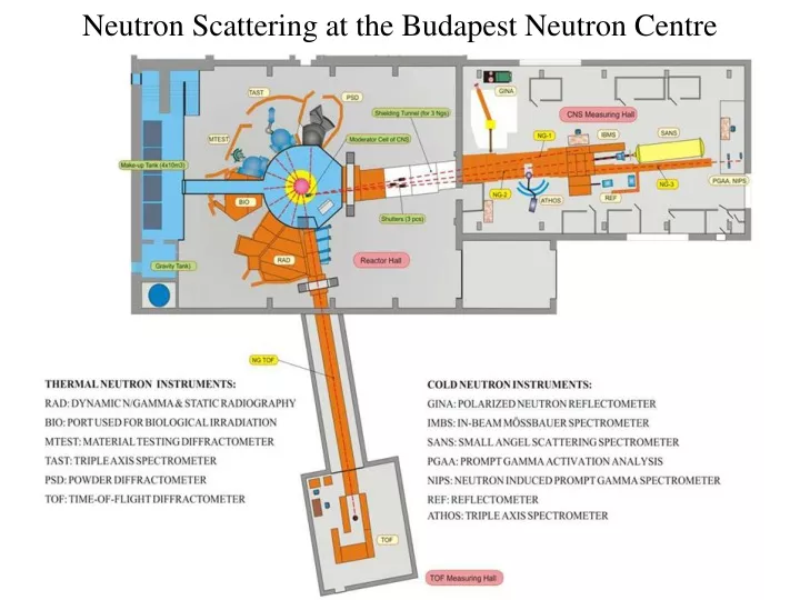
Neutron Scattering at the Budapest Neutron Centre. MTEST: Material Testing Diffractometer. The Ordela 1200N position sensitive detector has been installed at the MTEST diffractometer. Actually it is in regular operation. Detector in “far” position.

E N D
MTEST: Material Testing Diffractometer The Ordela 1200N position sensitive detector has been installed at the MTEST diffractometer. Actually it is in regular operation. Detector in “far” position Total (Bragg and diffuse) scattering measurements on powder, liquid and amorphous materials. A four-circle goniometer provides conditions for texture measurements. Ni pattern recorded with 1.45A° neutrons in “near” configuration
PSD: PowderDiffractometer Amorphous materials borosilicate glasses: suitable materials for the storage of high-level radioactive waste materials boromolybdate glasses:important physical and chemical properties, large ion- and electron conductivity, catalytic activity, non-linear optical properties, mechanical resistance mixed alkali silicate-bioactiveglasses: in contact with body fluids, glasses induce a specific biological response at their surface chalcogenide glasses: wide area of application as electrical and optical components, semiconductors...
PSD: PowderDiffractometer New in-situ sample environment high temperature pressure cell has been developed. • Temperature and pressure interval: • TiZr cell: Tmax: 450°C pmax: 300 bar • Saphire cell: Tmax: 950°C pmax: 300 bar • Study of CO2 capture in supercritical conditions • - Neutron diffraction study of thermal phase stability (exp. Alloys) • - Neutron diffraction in temperature dependent deformation (crystallization, new texture) • - Neutron diffraction study on electrode materials (based on Li or Ni elements) • Characterization of nanostructured alloys in extreme environment
TOF: Time-of-FlightDiffractometer Installed on a radial thermal neutron beam in a new guide-hall. The main advantage of TOF monochromatization in neutron diffractometry on a continuous source is the variable resolution and intensity. A full diffraction spectrum can be gained within a variable bandwidth with ultrahigh resolution or high intensity at conventional resolutions.
TOF: Time-of-FlightDiffractometer New detector and data acquisition system has been installed and commissioned • Backscattering detector bank: • 2θ= 144°-165° • 72-88 3He tubes • Optimized for HR diffraction • Lower angle detector bank: • 2θ= 120°, 90°, 60° • 24 3He tubes • Extension of Q-range Common, List mode DAQ
TOF: Time-of-FlightDiffractometer Construction of the backscattering bank Tubesize: 300x30x15mm (squashed) Pressure of 3He: 3bar Efficiency: 75% at 4Å – 30% at 1Å 7 boxes in 3 rows, +1 Boxes can individually be aligned in two direction Tubes will be aligned within the box for time-focusing. The positions and the orientation of the tubes can easily be calculated - even if the bank is replaced. The lower angle bank is similar, but 2 boxes in 3 rows.
1 0 6 -1 5 x10 -2 4 -3 -3 3 2 -4 -5 -1 0 1 2 3 4 5 TOF: Time-of-FlightDiffractometer Optimization of the backscattering bank • Compromising between resolution and detector solid angle. • Aligning the tubes individually, Δd<2x10⁻³Å resolution can be kept for almost the entire detector bank. • Intensity gain > 10 • Solid angle: x 17 • Resolution improves: 1.5x10⁻³Å at higher angles. Colors: available Δd [Å] White lines: constant d(t) surfaces – time focusing Positions in m, the sample is in the origo.
TOF: Time-of-FlightDiffractometer Positioner for large samples • Precession and nutation about the beam axis are provided, rotation can be added later; • Nutaion frame plane offset to allow free access to and scattered beam extraction from the nutation axis; • Large free solid angle for backscattering experiments.
TOF Simulation of Wavelength Frame Multiplication on a Long Pulse Spallation Source Principle Proposed by F. Mezei and M. Russina, tested on the BNC TOF instrument by Gy. Káli.
GINA: Polarized Neutron Reflectometer Thinlayerinvestigations, includingmagneticlayers. Superconductor-ferromagnetinterface.
GINA: Polarized Neutron Reflectometer New four-bounce polarizer and analyzerfiltershavebeeninstalled Concept of the polarizer filter • Technicaldata • Polarization efficiency: 99,3 % • Transmission: 74,5% • Advantages • Extremely high polarizationefficiency • Maintenance-free • Optical axis conserved • Disadvantage • Relatively lowtransmission Picture of the setup inactualuseonthe GINA reflectometer
GINA: Polarized Neutron Reflectometer New sampleenvironmentcell has beendeveloped • Motivation: • Tostudysolid-liquid interfaces • Controlled temperature • Controlled liquid composition forcontrast enhancement • Current status: • Being tested • Background measurements • Heat stability • ContrastmatchingSi-wafer The existing setup
SANS: Small Angle Scattering Spectrometer The SANS diffractometer Yellow Submarine covers a Q-range from 0.003 to 0.7 Å-1 allowing to probe structures at length scales from 5 Å to 1500 Å. It has a wide range of applications from studies of defects and precipitates in materials, surfactant and colloid solutions, ferromagnetics, magnetic correlations, alloy segregation, polymers, proteins, biological membranes.
SANS: Small Angle Scattering Spectrometer On isolated plant thylakoid membranes we could identify a peak originating from domains of ordered, unappressed stroma lamellae (a part of the highly organized thylakoid membrane assembly), providing information about its averaged repeat distance (RD) values. We found this RD to depend on e.g. the osmolarity and the ionic strength of the suspension medium. We also determined characteristic RDs of thylakoid membranes in algal or cyanobacterial cells and correlated these RDs with the size and the arrangement of the different protein complexes in the thylakoid membranes. Time-resolved SANS measurements were performed on these samples and revealed light-induced reversible reorganizations in the seconds-to-minutes time scale, which appeared to be associated with functional changes in vivo. Effect of illumination on the two-dimensional SANS profile of magnetically oriented spinach thylakoid membranes, recorded with the 2D detector of the D22 SANS instrument (ILL). Dark-adapted state (A), after illumination with white light of 1700 μmol photons m-2 s-1 photon flux density for 5 min (B), after 5 min light and 5 min dark (C).
SANS: Small Angle Scattering Spectrometer SANS investigation of a duplex steel (containing two different phases) of industrial interest subjected to various ageing processes. We have observed the growth of cuboid particles with size changes from a ≈ 17 nm to a≈ 20.5 ± 6.6 nm due to thermal treatment. Further thermal treatment causes a developing of polydispersity of scattering particles. Therefore in this case only an average size was estimated. It was shown that this system becomes very polydisperse and the scattering data can be approximated by average parameter of particles as a ≈ 17 nm. SANS analysis of steel samples (a) S1 as received, (b) S2 aged at low temperature, (c) S3 aged at high temperature. This yields the characteristic effect ofthe thermal treatment and helps evaluate the aging process.
SANS: Small Angle Scattering Spectrometer Original Publication Ünnep R, Zsiros O, Solymosi K, Kovács L, Lambrev PH, Tóth T, Schweins R, Posselt D, Székely NK, Rosta L, Nagy G, Garab G. (2014) The ultrastructure and flexibility of thylakoid membranes in leaves and isolated chloroplasts as revealed by small-angle neutron scattering. Biochim Biophys Acta, 1837(9):1572-80.
FSANS high resolution extension of the BNC Yellow Submarine SANS instrument • Single beam pinhole collimator • transfer parameter: AW 1.5105 cm2sr; • angular resolution: 1.9 mrad; • optimal wavelength resolution: Dl/l 12%; • sample size: 10 mm. • Multibeam converging collimator • transfer parameter: nAW 3.8105 cm2sr; • angular resolution: 0.56 mrad; • optimal wavelength resolution: Dl/l 3.6%; • sample size: 28 mm. • Elliptical focusing • transfer parameter: AW 2.3105 cm2sr • angular resolution: 0.9 mrad; • optimal wavelength resolution: Dl/l 6%; • sample size: 60 mm.
FSANS Elliptical reflective focusing Nested Kirkpatrick-Baez mirror pair First beam test results
FSANS Test bench for optical developments
ATHOS: Cold Neutron Three-Axis Spectrometer A triple-axis spectrometer has been designed for structural and dynamical studies of condensed matter. It is used in a multi-purpose regime: high resolution diffractometry, strain analysis, quasielasctic and inelastic scattering.
TAST: Thermal Neutron Three-Axis Spectrometer The TAST instrument provides moderate resolution (~2.0 meV) with sufficient intensity for use in a wide range of problems. It is ideally suited for the study of phonon and magnon dispersion curves in single crystals, phonon density of states for large class of materials which contain hydrogen. The flexibility of the triple axis spectrometer enables to use it in a multi-purpose regime e.g. transmission measurements and spectrometer components tests are done also.
TAST: Thermal Neutron Three-Axis Spectrometer • Atomic resolution neutron holography • internal source method (ammonium-chloride) • internal detector (converter) method (lead-cadmium alloy) • neutron holography of policrystalline samples • holographic reconstruction of magnetic fields at atomic level 3D reconstruction of nuclei positions around H in PdH.
REF: Reflectometer New monochromatorshavebeeninstalled and tested – nowinregularoperation -PG with 0.4º mosaicity instead of 3º Special alignment of crystal elements for optimal neutron transportation and focusing to the sample centre -horizontal neutron SMs between slits on top and bottom Monte-Carlo simulations (VITESS): 14 times increased intensity
REF: Reflectometer Monochromatoralignment -optical alignment (using laser beam and a screen) of individual crystals (slightly diffuse reflection from PG surface) - crystal positions adjusted in neutron beam The measured yield of individual crystals of the outer monochromator. Symmetrical intensity distribution.