Download
1 / 53
530 likes | 1.13k Views
How Cells Are Put Together. Chapter 4. The Cell. Smallest unit of life Can survive on its own or has potential to do so Is highly organized for metabolism Senses and responds to environment Has potential to reproduce. Structure of Cells. Two types: Prokaryotic Eukaryotic .

E N D
How Cells Are Put Together Chapter 4
The Cell • Smallest unit of life • Can survive on its own or has potential to do so • Is highly organized for metabolism • Senses and responds to environment • Has potential to reproduce
Structure of Cells Two types: • Prokaryotic • Eukaryotic All start out life with: • Plasma membrane • Region where DNA is stored • Cytoplasm
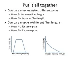
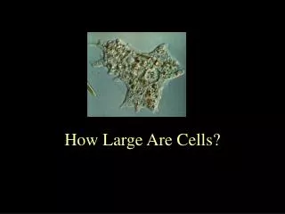
Why Are Cells So Small? • Surface-to-volume ratio • The bigger a cell is, the less surface area there is per unit volume • Above a certain size, material cannot be moved in or out of cell fast enough
Early Discoveries • Mid 1600s - Robert Hooke observed and described cells in cork • Late 1600s - Antony van Leeuwenhoek observed sperm, microorganisms • 1820s - Robert Brown observed and named nucleus in plant cells
Developing Cell Theory • Matthias Schleiden • Theodor Schwann • Rudolf Virchow
Cell Theory 1) Every organism is composed of one or more cells 2) Cell is smallest unit having properties of life 3) Continuity of life arises from growth and division of single cells
Microscopes • Create detailed images of something that is otherwise too small to see • Light microscopes • Simple or compound • Electron microscopes • Transmission EM or Scanning EM
Limitations of Light Microscopy • Wavelengths of light are 400-750 nm • If a structure is less than one-half of a wavelength long, it will not be visible • Light microscopes can resolve objects down to about 200 nm in size
Electron Microscopy • Uses streams of accelerated electrons rather than light • Electrons are focused by magnets rather than glass lenses • Can resolve structures down to 0.5 nm
incoming electron beam Electron Microscope condenser lens (focuses a beam of electrons onto specimen) specimen objective lens intermediate lens projector lens viewing screen (or photographic film)
Lipid Bilayer • Main component of cell membranes • Gives the membrane its fluid properties • Two layers of phospholipids
Fluid Mosaic Model • Membrane is a mosaic of • Phospholipids • Glycolipids • Sterols • Proteins • Most phospholipids and some proteins can drift through membrane
Membrane Proteins • Adhesion proteins • Communication proteins • Receptor proteins • Recognition proteins
Prokaryotic Cells • Archaea and eubacteria • DNA is not enclosed in nucleus • Generally the smallest, simplest cells • No organelles
Prokaryotic Structure bacterial flagellum pilus plasma membrane bacterial flagellum Most prokaryotic cells have a cell wall outside the plasma membrane, and many have a thick, jellylike capsule around the wall. cytoplasm, with ribosomes DNA in nucleoid region
Eukaryotic Cells • Have a nucleus and other organelles • Eukaryotic organisms • Plants • Animals • Protistans • Fungi
Functions of Nucleus • Keeps the DNA molecules of eukaryotic cells separated from metabolic machinery of cytoplasm • Makes it easier to organize DNA and to copy it before parent cells divide into daughter cells
Components of Nucleus Nuclear envelope Nucleoplasm Nucleolus Chromatin
Chromatin • Cell’s collection of DNA and associated proteins • Chromosome is one DNA molecule and its associated proteins • Appearance changes as cell divides
Nuclear Envelope • Two outer membranes (lipid bilayers) • Innermost surface has DNA attachment sites • Pores span bilayer one of two lipid bilayers (facing nucleoplasm) NUCLEAR ENVELOPE nuclear pore (protein complex that spans both lipid bilayers) one of two lipid bilayers (facing nucleoplasm)
Nucleolus • Dense mass of material in nucleus • May be one or more • Cluster of DNA and proteins • Materials from which ribosomal subunits are built • Subunits must pass through nuclear pores to reach cytoplasm
Endomembrane System • Group of related organelles in which lipids are assembled and new polypeptide chains are modified • Products are sorted and shipped to various destinations
Components of Endomembrane System Endoplasmic reticulum Golgi bodies Vesicles
Endoplasmic Reticulum • In animal cells, continuous with nuclear membrane • Extends throughout cytoplasm • Two regions: rough and smooth
Rough ER • Arranged into flattened sacs • Ribosomes on surface give it a rough appearance • Some polypeptide chains enter rough ER and are modified • Cells that specialize in secreting proteins have lots of rough ER
Smooth ER • A series of interconnected tubules • No ribosomes on surface • Lipids assembled inside tubules • Smooth ER of liver inactivates wastes, drugs • Sarcoplasmic reticulum of muscle is a specialized form
Golgi Bodies • Put finishing touches on proteins and lipids that arrive from ER • Package finished material for shipment to final destinations • Material arrives and leaves in vesicles
Vesicles • Membranous sacs that move through the cytoplasm • Lysosomes • Peroxisomes
Central Vacuole • Fluid-filled organelle • Stores amino acids, sugars, wastes • As cell grows, expansion of vacuole as a result of fluid pressure forces cell wall to expand • In mature cell, central vacuole takes up 50-90 percent of cell interior
Mitochondria • ATP-producing powerhouses • Double-membrane system • Carry out the most efficient energy-releasing reactions • These reactions require oxygen
Mitochondrial Structure • Outer membrane faces cytoplasm • Inner membrane folds back on itself • Membranes form two distinct compartments • ATP-making machinery is embedded in the inner mitochondrial membrane
Chloroplasts Convert sunlight energy to ATP through photosynthesis
Structure of a Chloroplast • Two outer membranes around semifluid interior (stroma) – bathes inner membrane • Often, this single membrane is folded back on itself as a series of stacked, flattened disks • Each stack is called a thylakoid, which contains chlorophylls and other substances involved in photosynthesis
Like Bacteria? • Both mitochondria and chloroplasts resemble bacteria • Have own DNA, RNA, and ribosomes
Cytoskeleton • Present in all eukaryotic cells • Basis for cell shape and internal organization • Allows organelle movement within cells and, in some cases, cell motility
Cytoskeletal Elements intermediate filament microtubule microfilament
Microtubules • Largest elements • Composed of the protein tubulin • Arise from microtubule organizing centers (MTOCs) • Polar and dynamic • Involved in shape, motility, cell division
Microfilaments • Thinnest cytoskeletal elements • Composed of the protein actin • Polar and dynamic • Take part in movement, formation and maintenance of cell shape
Accessory Proteins • Attach to tubulin and actin • Motor proteins • Crosslinking proteins
Intermediate Filaments • Present only in animal cells of certain tissues • Most stable cytoskeletal elements • Six known groups • Desmins, vimentins, lamins, etc. • Different cell types usually have 1-2 different kinds
Mechanisms of Movement • Length of microtubules or microfilaments can change • Parallel rows of microtubules or microfilaments actively slide in a specific direction • Microtubules or microfilaments can shunt organelles to different parts of cell
Flagella and Cilia • Structures for cell motility • 9 + 2 internal structure
False Feet • Some free-living cells, such as amoebas, form pseudopods (“false feet”) • These temporary, irregular lobes project from the cell and function in locomotion and prey capture • Pseudopods move as microfilaments elongate inside them – motor proteins attached to the microfilaments drag the plasma membrane with them
Cell Wall Plasma membrane • Structural component that wraps around the plasma membrane • Occurs in plants, some fungi, some protistans Primary cell wall of a young plant
Plant Cell Walls Secondary cell wall (3 layers) Primary cell wall
Plant Cuticle • Cell secretions and waxes accumulate at plant cell surface • Semi-transparent • Restricts water loss