Download
1 / 21
210 likes | 295 Views
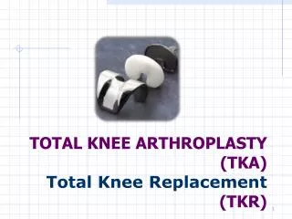
Comparison Between Computer-Assisted-Navigation and Conventional Total Knee Arthroplasties in Patients Undergoing Simultaneous Bilateral Procedures. A Randomised Clinical Trial Zhang G, Chen J, Chai W, Liu M, Wang Y, JBJS (2011)

E N D
Comparison Between Computer-Assisted-Navigation and Conventional Total Knee Arthroplasties in Patients Undergoing Simultaneous Bilateral Procedures A Randomised Clinical Trial Zhang G, Chen J, Chai W, Liu M, Wang Y, JBJS (2011) Stewart Morrison Western Health Orthopaedic Journal Club, July 2011
Background I • Many factors influence TKR longevity, including soft tissue balance, component design, post-operative complications, and accuracy of component alignment • Association between prosthetic alignment and TKR long-term survival has been well established • Jeffery et al (1991). • Within 3° of mechanical axis: 3% loosening at 12 years • Beyond 3° of mechanical axis: 24% loosening at 8 years
Background II • Computer navigation has been in clinical use for over 10 years • Conflicting evidence as to improvement of alignment • No improvement in alignment: Mielke (2001), Jenny (2001), Kim (2007) • Improvement in alignment: Bäthis (2004), Jenny (2003) • Increased operative time • Set out to compare computer navigated and conventional TKR, based upon • Component alignment • Operative time • Knee function • Performed in patients treated with simultaneous bilateral TKR, different technique used on each knee.
Method I Design Prospective Randomized Controlled Trial Minimum sample size of 32 subjects (64 knees) required, based on literature (contrast to Kim et al) Operative technique and order of operations randomised Inclusion criteria: candidate for bilateral cruciate retaining TKR Exclusion criteria: none stated Flexion Deformity: Computer Assisted Navigation 8.2° ± 2.9°, Conventional 8.3° ± 2.7° (p=0.882) Varus Deformity: Computer Assisted Navigation 7.8° ± 2.6°, Conventional 8.6° ± 2.5° (p=0.776) Link Gemini Mk II Prostheses BrainLab VectorVision CT-free Navigation
MethodII Medial Parapatellar Approach Conventional • Tibial anatomical axis defined as the line between the medial one-third of the tibial tubercle and a point 3 mm inward from the midpoint between the medial and lateral malleolus. 10 mm resection, 5° posterior slope. • Intramedullary instrumentation for femoral cuts. ER referenced off transepicondylar axis. ComputerAssisted • Bicortical femoral and tibial infrared trackers • Registration of center of rotation of femoral head, distal femur, proximal tibia, and medial and lateral malleoli. • Tibia cut with 10 mm resection, 5° posterior slope. • Femur cut orthogonal to the mechanical axis of the lower limb. ER based on transepicondylar axis. Routine soft tissue balancing, component implantation, and post operative rehabilitation.
MethodIII DataCollection • Full length standing XR • Cross Sectional CT of knee joints Measurements • Varus/Valgus angles of tibia (coronal plane) • Angle between horizontal axis of prosthesis and mechanical axis of limb (coronal plane) • Posterior slope tibial component (sagittal plane) • Flexion of femoral component (sagittal plane) • Rotational angle of femoral component relative to transepicondylar axis (axial CT) • Hospital for Special Surgery (HSS) scale • 6 month follow up by blinded surgeon
Results I Significant difference between groups with respect to: • Accuracy of tibial resection in both coronal and sagittal planes • Better approximation of the normal mechanical axis of the limb in coronal and sagittal plane In favour of computer-navigation group. Scatter of data on coronal plane smaller in computer navigated group Coefficient of variation of data in conventional group 3 x that of computer navigated group. Axial Alignment >3° deviation in 9 (28%) “conventional” knees, 0 (0%) CN knees Tibial component deviation from Tibial axis >3° 4 (13%) “conventional” knees, 0 (0%) CN knees
Results II Significant differences with respect to posterior slope, but actual difference and coefficient of variation small. No significant difference with regards to external/internal rotation, between groups. No significant difference in increase in HSS score at 6 months, between groups (p=0.0956) No pin site fracture or infection in CA group Operative time 30 min longer in CA group
Discussion “Our study demonstrated that TKR with computer-assisted navigation provided more accurate bone alignment in both the coronal and sagittal plane” “There was no significant difference between the two groups in terms of femoral rotational alignment or early functional recovery” No notching of femur in computer-navigated group
Discussion II Why is this important? • Sagittal plane alignment along mechanical axis to facilitate knee extension/avoid overextension • This also lessens wear in posterior-cruciate substituting prostheses
Discussion III “Because of variation of medial and lateral femoral condyles used for rotational alignment, and the accuracy of intraoperative marking of these structures, computer assisted navigation cannot ensure the accuracy of bone resection with external rotation” Potential Complications • Pin site fracture (large pin diameter, improper position, repeated drilling, obesity, osteoporosis, post operative trauma) • Pin track infection • Increased operative time
Discussion IV Prospective, self-controlled, randomised, single blind trial Limitations • Failure to compare difference in systemic complications cause by two techniques • Single, CR prosthesis • 6 month follow up only • No exclusion criteria given • No measure of relative severity of knee deformity or OA • No long term functional outcome data • No long term revision rate data • No comment as to the alignment of the axial CT slices (? Orthogonal to mechanical or anatomical axis of limb, or axis of torso)
“It is difficult for the patient with bilateral TKRs to separate the function of each knee”- Kim et al (2007)
Where to now? Review of local practices Meta-analysis • Zhang et al (2010) – 32 bilateral TKRs • Kim et al (2007) – 100 bilateral TKRs • Jenny et al (2004) – 40 + 40 TKRs • Bäthis et al (2004) – 80 + 80 TKRs • Kim et al (2003) – 555 TKRs • Mielke et al (2001) – 60 TKRs (not randomised) Functional outcome data Revision data